Women's Health
What Is a Normal Testosterone Level in Perimenopause?
Testosterone decline in perimenopause is often overlooked — yet it drives fatigue, brain fog, and low libido before estrogen levels even shift noticeably. Understanding what 'normal' actually means for your decade, and why free testosterone often matters more than total, is the first step toward reclaiming energy and clarity.
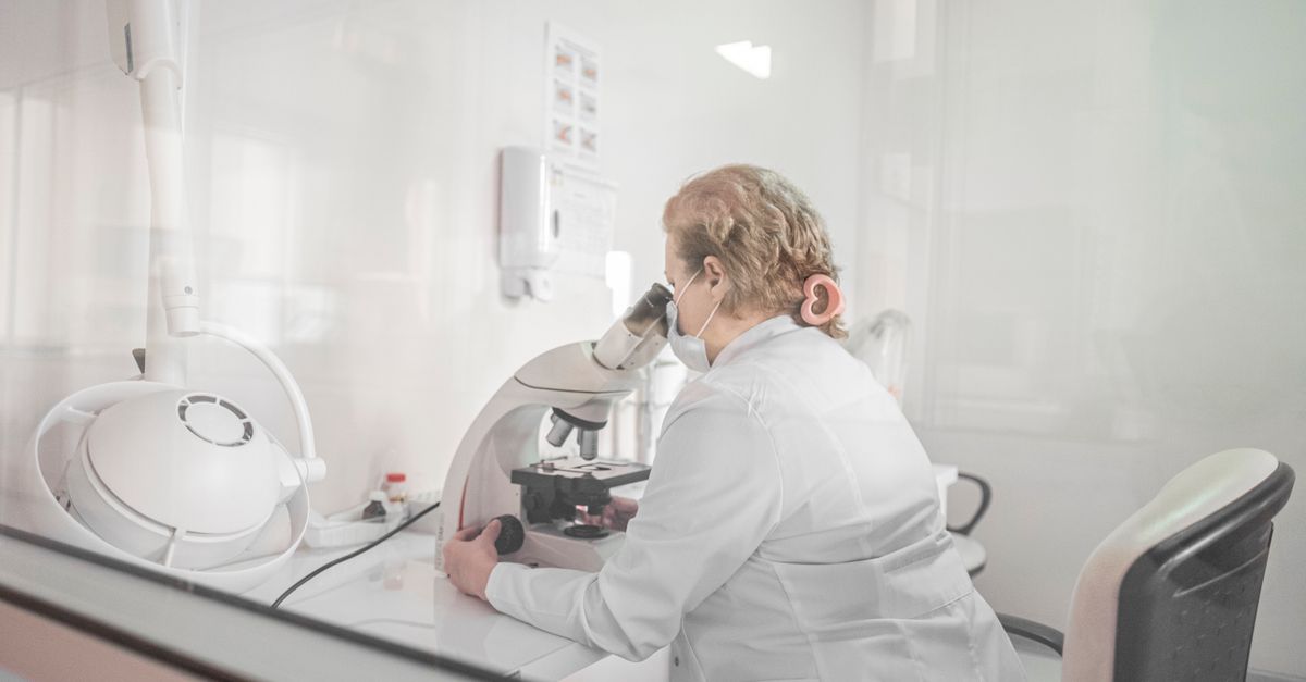
What Is a Normal Testosterone Level in Perimenopause?
For most women in perimenopause, a total testosterone level between 15–70 ng/dL is considered within the broad reference range, but "normal" is not the same as optimal. Many women feel their best in the upper third of that range, roughly 40–70 ng/dL. The key caveat: numbers mean little without free testosterone and SHBG context, because binding proteins can make adequate total testosterone functionally useless.
---
Why Testosterone Matters More Than Most Women Are Told
Testosterone is commonly framed as a male hormone, but women produce it in meaningful quantities — roughly 10–25% of what men produce — and it plays a central role in energy metabolism, mood regulation, bone density, libido, and cognitive sharpness. In perimenopause, ovarian testosterone output declines alongside estrogen, often starting in the mid-30s and accelerating through the 40s (Davison et al., Journal of Clinical Endocrinology & Metabolism 2005; PMID: 15827095).
What makes this particularly important is timing: testosterone can begin falling before estrogen does, which means classic perimenopause symptoms — unexplained fatigue, flat mood, difficulty concentrating, reduced strength — may be driven in part by androgens, not just estrogen. If only estrogen gets measured and managed, half the hormonal picture is invisible.
Testosterone also exerts direct effects on mitochondrial biogenesis and oxidative phosphorylation efficiency in skeletal muscle. When androgen receptor signaling declines, cells produce less ATP per unit of substrate — a cellular-level explanation for the disproportionate fatigue that many perimenopausal women report even with normal thyroid panels and adequate sleep. A 2019 review in Frontiers in Endocrinology noted that testosterone's role in mitochondrial function is increasingly recognized as independent of its anabolic effects on muscle mass itself (Varlamov et al., Frontiers in Endocrinology 2019; PMID: 30972036).
Symptoms commonly linked to low testosterone in perimenopausal women include:
- Persistent low energy that doesn't resolve with adequate sleep
- Reduced motivation or blunted sense of reward
- Decreased libido and reduced physical sensation
- Difficulty maintaining muscle mass despite regular exercise
- Brain fog and word-finding difficulty
- Unexplained exhaustion in perimenopause that lab panels often miss
Recognizing these as potentially androgen-related — rather than purely stress or estrogen-related — opens up more targeted options.
---
Total Testosterone Normal Range by Age
Reference ranges for women's testosterone vary between labs and assay methods, which creates genuine confusion. The table below reflects commonly used ranges, but your lab's specific reference interval should always be checked.
| Age Range | Broad Reference Range (ng/dL) | Functional Optimal Zone (ng/dL) |
|---|---|---|
| 20–29 | 20–75 | 40–75 |
| 30–39 | 15–70 | 35–70 |
| 40–49 (perimenopause) | 10–55 | 30–55 |
| 50–59 (early postmenopause) | 7–40 | 20–40 |
| 60+ | 5–30 | 15–30 |
Note that the "low normal" end of each decade's range corresponds closely with symptom burden in clinical cohorts. A woman at 42 with a total testosterone of 12 ng/dL is technically within some labs' reference range but is likely to report significant androgen-related symptoms (Davis et al., The Lancet Diabetes & Endocrinology 2015; PMID: 26358173).
It's also worth knowing that testosterone peaks in the early morning and can vary by 20–40% across the day, so testing should ideally happen between 7–10 AM in a fasted or lightly fasted state for the most reproducible result.
Assay method also matters substantially. Immunoassay-based total testosterone tests — the most common in routine bloodwork — can be inaccurate at the low concentrations typical in women. Liquid chromatography-tandem mass spectrometry (LC-MS/MS) is the gold standard for female testosterone measurement. If your result is borderline or inconsistent with your symptoms, requesting LC-MS/MS specifically is worth discussing with your provider. For a broader view of how reference ranges shift with age across sex hormones, testosterone optimal ranges by age provides useful context for both men and women.
---
Free Testosterone Normal Range by Age
Total testosterone tells you how much is circulating, but free testosterone — the unbound fraction available to tissues — tells you how much is actually working. In perimenopause, sex hormone-binding globulin (SHBG) commonly rises, which binds more testosterone and lowers bioavailable levels even when total testosterone looks adequate.
For women, free testosterone is typically measured as a percentage of total or as an absolute value in pg/mL:
| Age Range | Free Testosterone (pg/mL) | % of Total |
|---|---|---|
| 20–29 | 1.0–8.5 | ~1.5–3.0% |
| 30–39 | 0.8–7.0 | ~1.2–2.5% |
| 40–49 | 0.5–5.0 | ~1.0–2.0% |
| 50–59 | 0.3–3.5 | ~0.8–1.8% |
| 60+ | 0.2–2.5 | ~0.7–1.5% |
A woman with total testosterone of 45 ng/dL but SHBG of 120 nmol/L may have free testosterone below 0.8 pg/mL — functionally deficient despite adequate total levels. This is why pairing testosterone with an SHBG assessment is essential for a complete hormonal picture.
Calculated free testosterone (using the Vermeulen formula based on total testosterone, SHBG, and albumin) is often more reliable than direct free testosterone immunoassays, which can be imprecise at low concentrations (Vermeulen et al., Journal of Clinical Endocrinology & Metabolism 1999; PMID: 10523012). Most specialist endocrinologists and functional medicine practitioners now prefer the calculated method, especially when direct assay results seem inconsistent with symptoms.
It is also worth noting that albumin-bound testosterone — which accounts for roughly 40–50% of circulating testosterone in women — is partially bioavailable, since albumin binds testosterone with lower affinity than SHBG does. The "bioavailable testosterone" figure (free + albumin-bound) is therefore sometimes a more relevant clinical target than free testosterone alone, particularly in women with low albumin from poor nutrition or chronic inflammation.
---
What Causes Testosterone to Drop in Perimenopause?
Several mechanisms converge during perimenopause to reduce androgen availability:
- Declining ovarian function — The theca cells of the ovary produce roughly 25% of circulating testosterone. As follicular reserve depletes, this output falls.
- Adrenal DHEA reduction — DHEA, the primary precursor to both testosterone and estrogen, peaks around age 25 and declines roughly 10% per decade. By perimenopause, DHEA-S levels may be 50% of their youthful peak (Labrie et al., Journal of Steroid Biochemistry and Molecular Biology 2003; PMID: 14623516).
- Rising SHBG — Estrogen fluctuations and insulin sensitivity changes during perimenopause can elevate SHBG, binding more of the available testosterone.
- Chronic stress and elevated cortisol — The hypothalamic-pituitary-adrenal (HPA) axis competes with the HPG (reproductive) axis. Prolonged cortisol elevation suppresses LH pulsatility and reduces androgen synthesis — a mechanism sometimes described as "cortisol steal," though that term oversimplifies the biochemistry.
- Thyroid dysfunction — Both hypothyroidism and subclinical thyroid changes — common in perimenopausal women — alter SHBG levels and downstream testosterone availability. Checking thyroid markers alongside sex hormones is clinically prudent.
Insulin resistance, which often worsens through the perimenopausal transition, adds another layer of complexity. Elevated insulin lowers SHBG — which sounds beneficial — but simultaneously drives androgen excess in the ovarian stroma, creating a paradoxical state where total testosterone may appear normal or even elevated while free testosterone distribution and receptor sensitivity are disrupted. This is the underlying mechanism that links PCOS-pattern androgen dysregulation with the perimenopausal metabolic shift, and why understanding estradiol context in hormonal conditions can sharpen interpretation even outside a formal PCOS diagnosis.
---
How to Interpret Your Results: Total vs. Free vs. Bioavailable
Understanding the difference between total and free testosterone measurements is critical before drawing conclusions from a single number. Here's a practical framework:
Step 1 — Total testosterone
If below 20 ng/dL in a symptomatic woman under 55, investigate further. If above 70 ng/dL, consider whether androgens are being over-produced (PCOS, adrenal tumor — rare).
Step 2 — SHBG
If SHBG is above 80 nmol/L, significant binding is likely reducing free testosterone regardless of total levels.
Step 3 — Calculated free testosterone
Using the Vermeulen formula, values below 0.5 pg/mL in symptomatic women warrant clinical attention.
Step 4 — DHEA-S
Low DHEA-S (below 75 µg/dL in perimenopausal women) suggests declining adrenal androgen production and may be a more modifiable target than testosterone directly.
Step 5 — Context
Symptoms, not numbers alone, drive clinical decisions. Two women with identical lab panels may have vastly different symptom experiences depending on receptor sensitivity, sleep quality, nutritional status, and stress load.
---
The Role of Stress in Testosterone Decline
Chronic psychological and physiological stress is one of the most consistent drivers of hormonal disruption in perimenopausal women, yet it's routinely underestimated in clinical settings. Cortisol and testosterone have an inverse relationship in multiple studies: acute stress transiently raises testosterone, but chronic HPA activation suppresses it (Mehta & Josephs, Hormones and Behavior 2010; PMID: 20026051).
The mechanistic pathway runs through two parallel routes. First, elevated glucocorticoids directly inhibit GnRH pulsatility in the hypothalamus, which reduces downstream LH secretion and thereby reduces the thecal cell stimulation that drives ovarian testosterone synthesis. Second, chronic cortisol elevation upregulates 5-alpha reductase activity in peripheral tissues, accelerating the conversion of testosterone to dihydrotestosterone (DHT) — a potent androgen that does not feed back positively on energy or mood in the way testosterone does. The net result is lower circulating testosterone alongside higher peripheral DHT, a hormonal signature that can produce symptoms of both androgen deficiency and androgen excess simultaneously.
Practical strategies that have clinical support for reducing stress-related hormone disruption include:
- Adaptogenic herbs — Ashwagandha (KSM-66, 600 mg/day) has demonstrated significant reductions in cortisol (28% vs. placebo in an 8-week RCT of 64 adults with chronic stress) and improvements in perceived stress scores. Reductions in HPA-axis overactivity may create a permissive environment for testosterone recovery (Chandrasekhar et al., Indian Journal of Psychological Medicine 2012; PMID: 23439798).
- Sleep prioritization — A single week of sleep restriction to 5 hours per night reduced testosterone in young men by 10–15% in one controlled study; similar mechanisms likely operate in women, given shared HPG architecture.
- Resistance training — 2–3 sessions per week of progressive resistance exercise is among the most evidence-backed strategies for supporting androgen levels and DHEA production in middle-aged women.
- Magnesium adequacy — Magnesium deficiency activates the HPA axis and amplifies the cortisol response to stress. Correcting deficiency supports both stress resilience and downstream hormone production.
- Reducing inflammatory load — Systemic inflammation, driven by poor diet, dysbiosis, or unresolved infections, suppresses the HPG axis. This is why comprehensive metabolic work-up matters more than chasing a single hormone number.
If you're also experiencing disrupted sleep patterns like waking at 3 AM, the cortisol-testosterone interaction becomes even more relevant — nighttime cortisol spikes are a key driver of both sleep disruption and androgen suppression.
The exception to the stress-suppression pattern is worth noting: women with underlying hyperandrogenism (elevated baseline androgens due to PCOS or adrenal hyperplasia) may maintain higher testosterone under stress because the adrenal contribution to androgen synthesis is preserved or even amplified by cortisol-driven ACTH stimulation. In these women, chronic stress paradoxically sustains androgen output while still disrupting reproductive cycle regularity. If your total testosterone is above 70 ng/dL alongside irregular cycles and stress symptoms, a different clinical investigation is warranted than the one described for low-androgen perimenopause.
---
What This Means for Your Formula
If your lab work or wearable data points to suboptimal testosterone, low DHEA-S, elevated cortisol, or high SHBG, a targeted nutritional strategy can meaningfully support androgen balance — even before considering clinical hormone therapy.
Here's how Ones approaches this based on your biomarker profile:
Ashwagandha KSM-66 (600 mg) — Ones uses the full clinical dose of KSM-66 standardized extract, the same form tested in the Chandrasekhar 2012 RCT that showed a 28% reduction in serum cortisol and a 44% reduction in perceived stress scores across 64 participants over 8 weeks. Reducing chronic cortisol burden is one of the most modifiable levers for preserving androgen production in perimenopause.
Adrenal Support System Blend — Ones' proprietary Adrenal Support blend is formulated to address HPA axis dysregulation — a root-level driver of both testosterone decline and fatigue in perimenopausal women. Rather than supplementing testosterone directly (which requires clinical prescription), supporting the adrenal precursor pathway (DHEA → testosterone) through adaptogenic and nutritional co-factors is a safer, evidence-aligned approach.
Zinc — Zinc is an essential cofactor in testosterone biosynthesis and 5-alpha reductase regulation. Deficiency is associated with lower total and free testosterone across age groups. Ones includes zinc in bioavailable form at doses calibrated to your bloodwork, since supplementing zinc when replete adds no benefit — and excess zinc can impair copper absorption.
Your Ones formula is built from your actual lab data, not a generic women's health stack. The AI evaluates your testosterone, SHBG, DHEA-S, cortisol markers, and thyroid function together to determine which ingredients — and at what doses — are warranted for your specific pattern.
---
Key Takeaways
- A total testosterone level of 15–70 ng/dL covers the broad reference range for women in perimenopause, but most women feel optimal in the upper half (30–55 ng/dL for the 40s decade).
- Free testosterone and SHBG must be assessed alongside total testosterone — elevated SHBG can render adequate total testosterone functionally low, and albumin-bound testosterone adds further nuance.
- Assay method matters: LC-MS/MS is the gold standard for women's testosterone measurement; standard immunoassay results at low concentrations should be interpreted cautiously.
- Testosterone often begins declining in the mid-30s, before estrogen shifts become significant, making androgen assessment an early and important part of perimenopausal evaluation.
- Chronic stress drives testosterone suppression through two parallel pathways — reduced GnRH/LH pulsatility and upregulated 5-alpha reductase — making stress management a direct hormonal intervention, not just a lifestyle suggestion.
- Symptoms in context — not numbers in isolation — should drive clinical decisions; always work with a healthcare provider when interpreting hormone panels or considering hormone therapy.