Supplements
What Causes Dry Eyes with Adenomyosis?
Dry eyes may seem unrelated to a uterine condition, but adenomyosis drives systemic hormonal and inflammatory changes that directly affect tear production. Understanding the connection can help you address both problems at the root rather than treating them as separate issues.
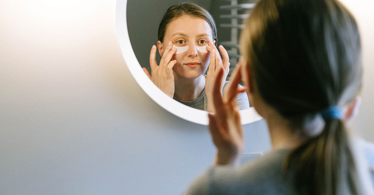
What Causes Dry Eyes with Adenomyosis?
Adenomyosis does cause dry eyes in many women, though the link is indirect. The same estrogen dominance, chronic low-grade inflammation, and omega-3 depletion that drive the uterine condition also impair the meibomian glands and reduce aqueous tear production. Women with stable hormone levels and no inflammatory burden rarely see this crossover.
Why Adenomyosis Affects the Eyes at All
Adenomyosis is defined by endometrial-like tissue growing into the myometrium, but its effects are not confined to the pelvis. Research consistently shows that women with adenomyosis have significantly elevated systemic markers of inflammation — including interleukin-6 (IL-6), tumor necrosis factor-alpha (TNF-α), and prostaglandin E2 — compared to age-matched controls (Benagiano et al., Human Reproduction Update 2012; PMID: 22313076). These same inflammatory mediators are expressed in the lacrimal gland and meibomian gland epithelium, where they suppress lipid secretion and aqueous tear output.
The ocular surface is not a sealed compartment. It is continuously bathed in secretions regulated by sex hormones — primarily estrogen, androgens, and prolactin. When systemic hormonal signaling is disrupted, as it is in adenomyosis, tear film stability deteriorates even though the eyes themselves are structurally normal. This explains why ophthalmologists often find no anatomical cause for dry eye in these patients, yet the symptom is real and often debilitating.
Similar hormone-ocular pathways appear across estrogen-related conditions. If you are also navigating dry eyes linked to hormonal shifts during perimenopause, many of the same mechanisms are at work.
The Estrogen Dominance Pathway
In adenomyosis, ectopic endometrial tissue is both estrogen-dependent and locally estrogen-producing. The lesions express aromatase — the enzyme that converts androgens to estrogens — at elevated levels, creating a microenvironment of persistent high estrogen even when serum estrogen looks within range on a lab panel (Bulun et al., Endocrine Reviews 2009; PMID: 19789222). Over time this shifts the systemic estrogen-to-progesterone ratio in a way that affects every tissue with sex hormone receptors, including the ocular surface.
Estrogen has a biphasic relationship with the lacrimal gland. At physiological levels it supports tear secretion. At chronically elevated or fluctuating levels, particularly when progesterone is relatively low, lacrimal gland function becomes erratic. Androgens — especially dihydrotestosterone (DHT) — are the primary trophic hormones for meibomian glands, which produce the oil layer that prevents tear evaporation. Estrogen dominance suppresses androgen signaling, reducing meibomian gland output and accelerating evaporative dry eye.
This same estrogen-androgen imbalance can also thin and dry the skin, a symptom frequently reported alongside dry eyes. Women experiencing dry skin alongside adenomyosis often share overlapping hormonal drivers with those reporting ocular dryness — both tissues are androgen-sensitive.
Prostaglandins, Inflammation, and the Tear Film
Adenomyosis lesions produce large quantities of prostaglandin E2 (PGE2) and prostaglandin F2α (PGF2α), partly explaining the severe dysmenorrhea characteristic of the condition. Systemically, chronically elevated prostaglandins have well-documented effects on the ocular surface: they increase vascular permeability in the conjunctiva, promote mast cell degranulation, and drive neurogenic inflammation in the corneal epithelium (Massingale et al., Cornea 2009; PMID: 19337156). The result is a tear film that is not only reduced in volume but also chemically destabilized — elevated matrix metalloproteinase-9 (MMP-9) in the tear fluid degrades mucin, shortening tear break-up time.
Women with adenomyosis who also have systemic histamine excess — itself driven by estrogen, since estrogen upregulates histidine decarboxylase — may experience an additional layer of ocular irritation. Histamine directly stimulates conjunctival goblet cell mucus secretion while simultaneously destabilizing the lipid layer, creating paradoxical symptoms: eyes that water excessively but still feel dry. If you notice other hormonal symptoms like premenstrual mood shifts or skin reactivity, dry eyes that worsen before your period may be part of the same histamine-estrogen loop.
Nutrient Depletion as a Contributing Factor
Chronic inflammation is metabolically expensive. Women with adenomyosis frequently present with depletion of anti-inflammatory micronutrients, particularly omega-3 fatty acids (EPA and DHA), vitamin D3, magnesium, and zinc — all of which play documented roles in tear film health.
Omega-3 fatty acids are incorporated directly into meibomian gland secretions and reduce lacrimal gland inflammation via competitive inhibition of arachidonic acid metabolism. A randomized controlled trial of 264 patients with dry eye disease found that omega-3 supplementation (EPA 2000 mg + DHA 1000 mg daily for 12 months) significantly improved tear break-up time and Schirmer test scores compared to placebo (Bhargava et al., Contact Lens and Anterior Eye 2013; PMID: 23218243).
Vitamin D3 receptors are expressed on lacrimal gland acinar cells, and vitamin D deficiency is independently associated with aqueous-deficient dry eye. A 2017 cross-sectional study found that serum 25(OH)D levels below 20 ng/mL were significantly associated with reduced Schirmer test scores and higher Ocular Surface Disease Index (OSDI) scores (Karaca et al., Eye 2016; PMID: 27174382). Women with adenomyosis have disproportionately high rates of vitamin D insufficiency, partly because chronic inflammation accelerates 25(OH)D catabolism.
Zinc is required for retinol-binding protein synthesis in the liver, and retinol (vitamin A) is critical for goblet cell density on the conjunctival surface. Goblet cells produce the mucin layer of the tear film; when zinc-mediated vitamin A transport is impaired, goblet cell numbers decline and mucin production drops, leading to early tear film breakup.
The interconnected nature of these depletions means that fatigue, hair changes, and dry eyes can all appear together. Exhaustion that comes with adenomyosis often traces back to the same inflammatory drain on B vitamins, iron, and magnesium that also contributes to poor tear film stability.
The Nervous System Connection
Adenomyosis is increasingly recognized as a condition that sensitizes the central nervous system. Chronic pelvic pain remodels pain-processing circuits, elevating sympathetic nervous system tone and reducing parasympathetic output. This matters for dry eye because tear secretion — particularly the aqueous component from the lacrimal gland — is parasympathetically driven via the facial nerve (cranial nerve VII). Elevated sympathetic tone blunts this reflex arc, reducing basal tear secretion independent of hormone levels or inflammation.
Women with adenomyosis often report a cluster of symptoms that reflect autonomic dysregulation: poor sleep, difficulty recovering from exercise, cold hands and feet, and — dry eyes. These are not coincidental. The adrenal-sympathetic response to chronic pain and hormonal instability creates a feedback loop that simultaneously worsens pelvic symptoms and reduces lacrimal output.
This systemic stress response can also manifest as hair thinning, since elevated cortisol shifts hair follicles into a telogen phase — another example of how a single hormonal disruption expresses itself across multiple organ systems.
Diagnosing the Overlap: What to Look For
If you have diagnosed or suspected adenomyosis and are experiencing dry eyes, the following pattern of findings is clinically meaningful:
| Finding | Clinical Significance |
|---|---|
| Short tear break-up time (<10 seconds) | Evaporative dry eye; suggests meibomian gland dysfunction |
| Low Schirmer test (<10 mm/5 min) | Aqueous deficiency; suggests lacrimal gland suppression |
| Elevated serum hsCRP | Confirms systemic inflammatory burden |
| Low serum 25(OH)D (<30 ng/mL) | Independent dry eye risk factor; common in adenomyosis |
| Low omega-3 index (<4%) | Predicts poor meibomian gland function |
| Elevated estradiol with low progesterone | Supports estrogen dominance as mechanism |
| Elevated serum ferritin with low serum iron | Indicates inflammatory iron sequestration |
A full hormone panel, inflammatory markers, and a comprehensive metabolic panel will often reveal this pattern simultaneously. Treating dry eye in isolation with artificial tears addresses symptoms temporarily but does not correct the upstream drivers.
What This Means for Your Formula
Addressing dry eyes in the context of adenomyosis requires working upstream — reducing systemic inflammation, correcting nutrient deficits, and supporting hormonal balance rather than patching the ocular surface alone.
The Ones platform analyzes blood work, wearable data, and health history to identify which of these mechanisms is most active for an individual. Based on those findings, a custom capsule formula is assembled from clinically validated ingredients. For someone presenting with the adenomyosis–dry eye pattern, the most relevant actives typically include:
- Omega-3 (EPA/DHA): Ones sources pharmaceutical-grade EPA and DHA at combined doses calibrated to the individual's omega-3 index. The clinical benchmark for ocular benefit is EPA ≥ 1000 mg + DHA ≥ 500 mg daily, consistent with the Bhargava et al. (2013) RCT cited above.
- Vitamin D3 + K2 (MK-7): Ones pairs D3 with MK-7 to optimize both calcium metabolism and immune modulation. Doses are adjusted to the individual's baseline 25(OH)D, targeting the 40–60 ng/mL range associated with optimal lacrimal gland function.
- Zinc (as zinc bisglycinate): Included when retinol transport markers or dietary intake suggest shortfall, supporting both goblet cell maintenance and immune regulation relevant to adenomyosis-driven inflammation.
- Ones Histamine Support blend: For women in whom estrogen-driven histamine excess is identified as a contributor to ocular irritation and cyclical symptoms, this proprietary blend addresses the histamine-diamine oxidase (DAO) pathway directly.
Formulas are built around a 6- or 9-capsule daily plan determined by the AI based on the individual's findings — not self-selected by the user.
Key Takeaways
- Adenomyosis drives dry eyes through at least four converging mechanisms: estrogen dominance suppressing androgen-mediated meibomian gland function, chronic prostaglandin-driven lacrimal inflammation, depletion of key tear-film nutrients (omega-3, vitamin D3, zinc), and autonomic nervous system dysregulation from chronic pelvic pain.
- Estrogen dominance suppresses androgen signaling in meibomian glands, reducing the oil layer of the tear film and accelerating evaporation.
- Systemic prostaglandins and elevated MMP-9 in tear fluid destabilize the mucin layer and shorten tear break-up time — independent of structural eye pathology.
- Omega-3 deficiency, vitamin D insufficiency, and zinc depletion are common in adenomyosis and each independently impair different layers of the tear film.
- The sympathetic nervous system dominance associated with chronic pelvic pain directly reduces parasympathetic lacrimal secretion, adding an autonomic component to aqueous-deficient dry eye.
- Effective treatment addresses the upstream hormonal and inflammatory drivers — not just the ocular surface — and should be guided by actual lab data rather than population-average supplement stacks.