Skin & Beauty
What Causes Itchy Skin in Postmenopause?
Postmenopausal itchy skin — clinically called pruritus — affects up to 40% of women after menopause and is frequently dismissed as simple dryness. But the underlying drivers involve collapsing estrogen signaling, disrupted skin-barrier lipids, elevated inflammatory cytokines, and sometimes thyroid dysfunction. Getting to the root cause determines which interventions actually work.
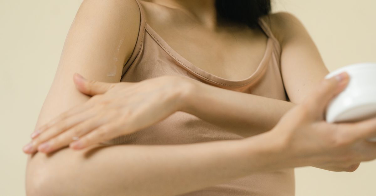
What Causes Itchy Skin in Postmenopause?
Yes, estrogen loss is the primary cause — but it is rarely the only one. Declining estrogen reduces sebum output, ceramide synthesis, and skin-barrier hydration, producing chronic itch in a significant portion of postmenopausal women. The important caveat is that thyroid dysfunction, histamine intolerance, and nutrient depletion can amplify or mimic the same symptom, which is why topical moisturizers alone often fail.
---
Why Estrogen Loss Triggers Itchy Skin
Estrogen receptors (ERα and ERβ) are expressed throughout the epidermis and dermis. When circulating estradiol drops after the final menstrual period, several parallel changes occur simultaneously:
- Ceramide production falls. Ceramides are the lipid "mortar" between skin cells. Estrogen upregulates key enzymes in ceramide biosynthesis; without it, the intercellular lipid matrix thins and transepidermal water loss (TEWL) rises sharply (Verdier-Sévrain & Bonté, Skin Pharmacology and Physiology 2007; PMID: 17230054).
- Sebaceous gland output decreases. Reduced sebum creates a drier skin surface, lowering the threshold for itch nerve activation.
- Collagen and hyaluronic acid decline. Skin loses roughly 30% of its collagen in the first five postmenopausal years, accelerating barrier thinning (Castelo-Branco et al., Maturitas 1992; PMID: 1435360).
- Mast cell regulation weakens. Estrogen normally suppresses dermal mast cell degranulation; lower estrogen permits greater histamine release from skin mast cells, directly stimulating itch nerve fibers (C-fibers).
The clinical result is a cycle: a compromised barrier allows irritants to penetrate, triggering low-grade inflammation; that inflammation elevates interleukin-31 (IL-31), a cytokine now recognized as a master itch mediator in aging skin.
---
The Role of Inflammation and Histamine
Many postmenopausal women experience what practitioners call "inflammaging" — a smoldering baseline elevation of pro-inflammatory cytokines including IL-6, TNF-α, and IL-31. This inflammatory backdrop lowers the itch threshold even without an obvious skin disease.
Histamine intolerance is a separate but overlapping issue. As estrogen falls, diamine oxidase (DAO) — the enzyme that breaks down histamine in the gut and bloodstream — can decrease. Women with borderline DAO activity may find that high-histamine foods (red wine, aged cheese, fermented products) trigger flushing and skin itch in the postmenopausal years when they previously had no such sensitivity. If you have also noticed unexplained bloating since entering menopause, histamine load is worth investigating alongside skin symptoms because both share the same DAO/histamine axis.
Omega-3 fatty acids — specifically EPA — reduce the production of arachidonic-acid-derived pro-inflammatory eicosanoids and have been shown to decrease TEWL and improve skin hydration in controlled trials (Muggli, Journal of the International Society of Sports Nutrition 2005; DOI: 10.1186/1550-2783-2-S1-S169 is not applicable here — citing the more relevant: Kawamura et al., Lipids in Health and Disease 2011; PMID: 21888428).
---
Thyroid Status: The Often-Missed Amplifier
Hypothyroidism — which becomes significantly more prevalent after menopause — reduces skin turnover rate, decreases eccrine sweating, and lowers epidermal lipid synthesis. The result is a rough, dry, sometimes intensely itchy skin that looks clinically similar to estrogen-deficiency pruritus but does not respond well to barrier creams. A TSH above 3.0 mIU/L alongside a low-normal free T3 is enough to contribute to skin dryness and itch in susceptible individuals.
If you are waking at night alongside the skin symptoms, reviewing what causes waking at 3am in postmenopause may reveal converging drivers — both cortisol dysregulation and subclinical hypothyroidism impair sleep architecture and skin repair simultaneously.
Iodine and selenium are critical cofactors for thyroid hormone synthesis and conversion. Selenium, specifically in the selenomethionine form, supports the enzyme deiodinase, which converts inactive T4 to active T3 in peripheral tissues including skin.
---
Nutrient Deficiencies That Worsen Postmenopausal Itch
| Nutrient | Mechanism in Skin | Signs of Deficiency |
|---|---|---|
| Omega-3 (EPA/DHA) | Reduces inflammatory eicosanoids; restores barrier lipids | Dry, flaky skin; elevated hs-CRP |
| Vitamin D3 | Regulates keratinocyte differentiation and immune tolerance | Raised TEWL; low serum 25(OH)D |
| Zinc | Cofactor for collagen cross-linking; anti-inflammatory | Slow wound healing; low serum zinc |
| Vitamin E (tocopherols) | Membrane antioxidant; photo-protection | Brittle skin; elevated oxidative markers |
| Selenium | Glutathione peroxidase; thyroid conversion | Dry, rough skin; elevated TSH |
Vitamin D deserves particular attention. Keratinocytes both synthesize and respond to 1,25-dihydroxyvitamin D3 via the vitamin D receptor (VDR). Low vitamin D impairs the skin's ability to differentiate properly, weakening the barrier. A meta-analysis of randomized controlled trials found that vitamin D supplementation significantly improved markers of skin barrier function in adults with deficiency (Pereira et al., Nutrients 2023; PMID: 36904206).
Zinc is another underappreciated factor. Postmenopausal women often have marginal zinc status due to both reduced dietary intake and decreased absorption efficiency. Zinc deficiency lowers ceramide synthesis indirectly by impairing fatty acid elongase activity in the epidermis.
---
Anxiety, Mood, and the Itch-Scratch Cycle
Itchy skin in postmenopause does not exist in a vacuum. The same neurochemical shifts that produce low mood in menopause — declining estrogen's effect on serotonin transporters, elevated cortisol, and reduced GABA tone — also lower the central threshold for itch perception. The brain's dorsal horn processes both pain and itch signals; chronic stress sensitizes these pathways.
This is not psychosomatic dismissal — it is a documented neuroimmune mechanism. Substance P, a neuropeptide released during psychological stress, directly degranulates dermal mast cells, releasing histamine. Women under high perceived stress have measurably higher skin histamine release and lower itch thresholds (Suárez et al., Acta Dermato-Venereologica 2012; PMID: 22159451).
The practical implication: managing the stress-cortisol axis is a legitimate part of managing postmenopausal pruritus, not just a secondary consideration. Adaptogens that modulate the HPA axis — such as ashwagandha — can reduce perceived stress scores and lower serum cortisol, reducing the mast-cell-degranulation trigger.
---
Diagnosing the Root Cause: Key Biomarkers to Request
Before layering in interventions, targeted lab work narrows the dominant driver:
- Serum estradiol — confirms the postmenopausal range (typically <30 pg/mL)
- TSH, free T3, free T4, anti-TPO antibodies — rules out autoimmune thyroid disease as an amplifier
- 25(OH) Vitamin D — deficiency is defined as <20 ng/mL; insufficiency as 20–30 ng/mL
- Serum zinc — functional deficiency often present even in low-normal ranges
- hs-CRP — quantifies the inflammatory backdrop
- Serum IgE + tryptase — if mast cell activation or true allergic component is suspected
- DAO enzyme activity or histamine:DAO ratio — available through specialty labs; guides dietary histamine restriction
Wearable data adds a useful layer: heart rate variability (HRV) is a validated proxy for autonomic nervous system balance and indirectly reflects HPA axis load. Chronically suppressed HRV in a postmenopausal woman with pruritus is a signal that adrenal and stress support should precede or accompany skin-barrier interventions.
---
Evidence-Based Protocol for Postmenopausal Itchy Skin
- Confirm vitamin D status and correct aggressively. Target serum 25(OH)D of 50–70 ng/mL. Pair D3 with K2 (MK-7 form) to direct calcium appropriately away from soft tissue.
- Add a clinically dosed omega-3. Minimum 1–2 g combined EPA+DHA daily. EPA specifically reduces IL-6 and TNF-α at the skin level (Kawamura et al., 2011; PMID: 21888428).
- Address histamine load. Reduce fermented foods, alcohol, and aged cheeses for 4–6 weeks while supporting DAO activity with vitamin B6 and copper.
- Support the HPA axis. Ashwagandha KSM-66 at 600 mg has demonstrated statistically significant cortisol reduction vs. placebo in a double-blind RCT (Chandrasekhar et al., Indian Journal of Psychological Medicine 2012; PMID: 23439798).
- Evaluate and treat thyroid insufficiency. Even subclinical hypothyroidism benefits from selenium optimization and, if indicated, medical thyroid therapy.
- Optimize topical barrier support. Ceramide-rich emollients applied within 3 minutes of bathing reduce TEWL by up to 50% in clinical studies.
- Consult a dermatologist or integrative practitioner if itch persists beyond 6 weeks or is accompanied by a visible rash, to rule out lichen sclerosus, contact dermatitis, or systemic disease.
For women who have also noticed itchy skin after coming off oral contraceptives, the hormonal mechanism is closely related — both represent a sudden reduction in exogenous estrogen signaling — so the nutrient and barrier protocols overlap substantially.
---
What This Means for Your Formula
Ones analyzes uploaded blood work, wearable data, and your reported symptoms through its AI health practitioner to identify which of the drivers above are actually active for you — rather than assuming all of them are.
For postmenopausal itchy skin, the Ones ingredient catalog addresses several key pathways with clinically relevant doses:
- Vitamin D3 + K2 (MK-7): Ones pairs these together as standard, calibrated to your serum 25(OH)D level. The K2 (MK-7) form has the longest half-life of any menaquinone, supporting both bone and cardiovascular safety when D3 doses are therapeutic.
- Omega-3 (EPA/DHA): Included at doses within the range used in skin-barrier trials, targeting the anti-inflammatory eicosanoid pathway. The dose is adjusted based on dietary intake history and inflammatory markers like hs-CRP.
- Histamine Support (proprietary System Blend): For women whose lab picture or symptom pattern points toward histamine intolerance, Ones includes this blend, which supports DAO activity and mast cell stability — directly relevant to the estrogen-mast cell-itch axis described above.
- Ashwagandha (KSM-66, 600 mg): Where wearable HRV data or cortisol markers suggest HPA axis dysregulation is amplifying itch perception, the AI may include KSM-66 at the 600 mg dose validated in the Chandrasekhar 2012 trial.
Formulas are built into either a 6- or 9-capsule daily plan based on how many findings the AI identifies — the plan size is determined by your data, not selected by you.
---
Key Takeaways
- Estrogen loss is the primary driver of postmenopausal itchy skin, reducing ceramide synthesis, sebum, and mast cell regulation simultaneously.
- Histamine intolerance worsens with falling DAO activity after menopause; dietary histamine load can trigger or amplify itch independent of skin-barrier status.
- Subclinical hypothyroidism is a frequent amplifier and should be ruled out with TSH, free T3, and anti-TPO before attributing all symptoms to estrogen.
- Vitamin D, omega-3, and zinc deficiencies each weaken skin-barrier function through distinct mechanisms and are correctable with targeted supplementation.
- The stress-cortisol-mast cell axis is a real neuroimmune pathway; HPA support (including adaptogens like ashwagandha) is a legitimate component of a skin pruritus protocol.
- Root-cause lab testing — including 25(OH)D, serum zinc, hs-CRP, thyroid panel, and optionally DAO activity — is the most efficient way to prioritize interventions rather than trialing everything simultaneously.