Minerals
What Does Iron Tell You About Your Hormones?
Iron is rarely discussed as a hormone regulator, yet it sits upstream of thyroid function, estrogen metabolism, and adrenal output. Even a 'normal' hemoglobin with a low-normal ferritin can quietly derail your hormones — and most standard panels miss it entirely.
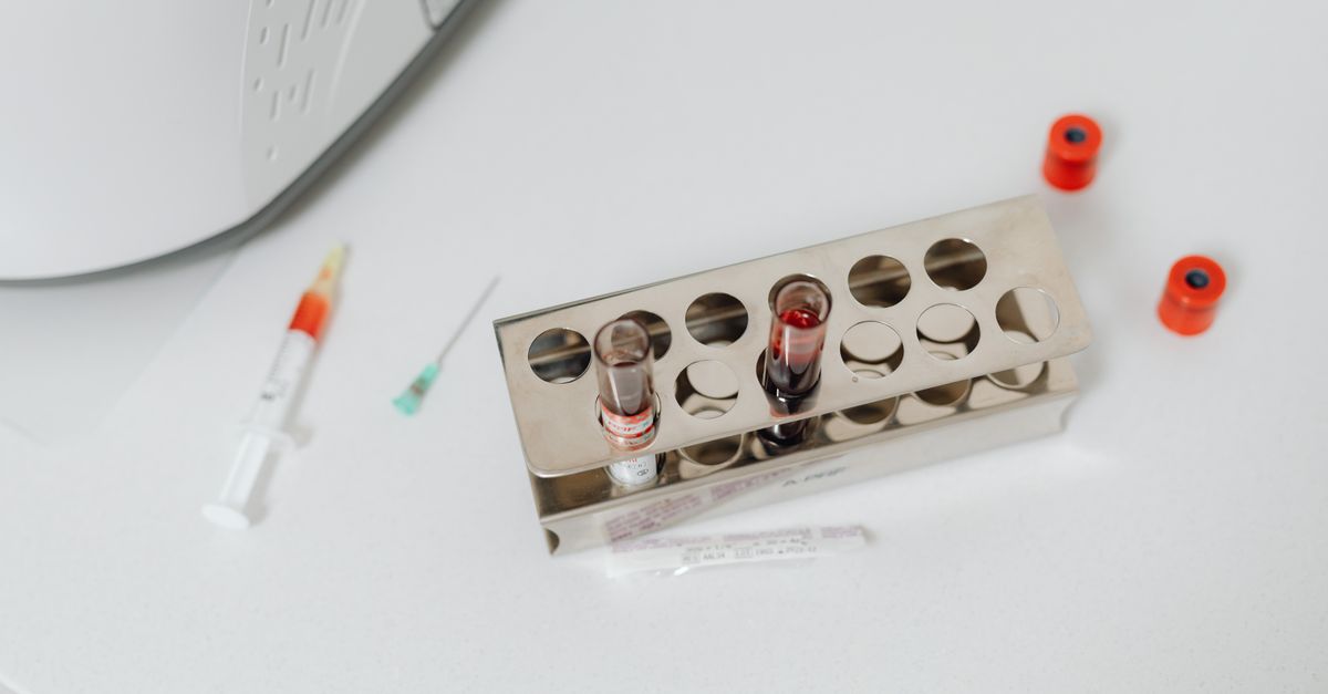
What Does Iron Tell You About Your Hormones?
Yes — quite a lot. Iron is a direct cofactor in thyroid hormone synthesis, estrogen clearance, and cortisol production, meaning suboptimal iron status can disrupt multiple hormonal axes simultaneously. The main caveat: standard CBC panels often miss early iron insufficiency. The exception is people with hemochromatosis, where excess iron suppresses hormones through a completely different mechanism.
---
Iron Is Not Just an Oxygen Carrier
Most people learn about iron in the context of anemia — low hemoglobin, pale skin, shortness of breath. That framing is accurate but incomplete. Iron functions as a metalloenzyme cofactor in dozens of enzymatic reactions, several of which sit at the center of hormonal regulation.
Here is the short list of where iron intersects with hormone physiology:
- Thyroid peroxidase (TPO) — the enzyme that converts iodide into thyroid hormones T3 and T4 — is a heme-iron-dependent enzyme. Without adequate iron, TPO activity drops and thyroid hormone output falls even when TSH looks normal.
- Cytochrome P450 enzymes (CYP450) — the liver's estrogen-metabolizing machinery — are heme-dependent. Low iron slows estrogen clearance and can contribute to estrogen dominance patterns.
- Dopamine beta-hydroxylase — required for converting dopamine to norepinephrine and ultimately supporting adrenal catecholamine output — also requires iron.
- Mitochondrial electron transport — iron-sulfur clusters in Complex I and Complex II drive ATP synthesis, which fuels every steroidogenic step in the adrenals and gonads.
So when you see borderline thyroid labs, unexplained cycle irregularities, or persistent fatigue alongside a low-normal ferritin, iron status deserves a serious look before assuming primary thyroid or hormonal pathology.
---
How Iron Deficiency Disrupts Thyroid Function Specifically
The thyroid-iron connection is one of the most clinically documented crossovers in micronutrient medicine. A systematic review published in Nutrients (Solberg et al., 2023; PMID: 37447305) confirmed that iron deficiency, even in the absence of overt anemia, significantly reduces thyroid peroxidase activity and impairs T4-to-T3 conversion.
In practical terms, this means:
- TSH may remain in range (typically 0.5–4.5 mIU/L) while free T3 is quietly low.
- Hypothyroid symptoms — cold intolerance, brain fog, slow metabolism, constipation — appear without a flagged TSH.
- Thyroid hormone replacement (levothyroxine) works less efficiently when iron is deficient, because cellular uptake of T3 depends partly on iron-containing mitochondrial enzymes.
A landmark intervention trial in Moroccan schoolchildren with combined iodine and iron deficiency showed that correcting iron deficiency alone improved thyroid function biomarkers significantly — more than iodine supplementation alone (Zimmermann et al., American Journal of Clinical Nutrition 2000; PMID: 10871577). While that population is pediatric, the enzyme biology is identical in adults.
For a deeper look at how TSH alone can mislead you, see what TSH tells you about your hormones.
---
Ferritin: The Number Your Doctor May Not Prioritize
Ferritin is the storage form of iron and the single most sensitive early marker of iron insufficiency — long before hemoglobin drops. The conventional lab range for ferritin is typically 12–300 ng/mL in women and 12–400 ng/mL in men. A value of 14 ng/mL is technically "normal" but functionally inadequate for many people.
Functional medicine practitioners and several research groups have proposed that optimal ferritin for hormonal and cognitive function sits between 50–100 ng/mL, particularly for premenopausal women.
| Ferritin Range (ng/mL) | Likely Iron Status | Common Hormonal/Symptomatic Correlates |
|---|---|---|
| < 15 | Depleted stores | Fatigue, hair shedding, restless legs, low T3 |
| 15–30 | Low-normal | Subclinical hypothyroid symptoms, poor exercise recovery |
| 30–70 | Adequate (general) | Most adults function well |
| 70–100 | Optimal (functional) | Preferred range for thyroid conversion, fertility, cognition |
| > 200 (unexplained) | Possible excess or inflammation | May signal hemochromatosis or inflammatory burden |
Importantly, elevated ferritin does not always mean iron excess — it is also an acute-phase reactant that rises with inflammation. This is why interpreting ferritin alongside CRP and transferrin saturation matters. To understand how CRP fits into this picture, see what CRP tells you about your hormones.
---
Iron, Estrogen, and the Menstrual Cycle: A Two-Way Relationship
Iron and estrogen influence each other in both directions, creating a feedback loop that is particularly relevant for reproductive-age women.
Estrogen upregulates iron absorption. Research published in Blood (Ikuta et al., 2012; PMID: 22234690) demonstrated that estrogen suppresses hepcidin — the liver hormone that limits iron absorption — effectively increasing intestinal iron uptake during the follicular phase of the cycle. This makes biological sense: iron is needed for the proliferating endometrium.
Iron deficiency disrupts estrogen metabolism. Cytochrome P450 1A2 (CYP1A2) and CYP3A4, both heme-iron-dependent enzymes, handle estrogen hydroxylation and conjugation in the liver. When iron is low, these pathways slow, and estrogen metabolites recirculate rather than being excreted. Clinically, this can look like:
- Heavier or longer periods (which then worsen iron loss — a vicious cycle)
- Premenstrual bloating and mood changes consistent with estrogen excess
- Fibrocystic breast tissue patterns
Menstrual blood loss accounts for roughly 15–20 mg of iron per period in a typical cycle, and up to 250 mg in women with heavy menstrual bleeding (Hallberg et al., Acta Obstetricia et Gynecologica Scandinavica 1966; PMID: 5968911). This foundational figure explains why iron insufficiency is disproportionately common in premenopausal women and why cycle irregularities and iron status should always be evaluated together.
This hormonal interplay is also connected to cortisol and adrenal function — a topic explored in detail at what does ferritin tell you about your hormones.
---
Iron and Adrenal / Cortisol Regulation
The adrenal connection is less discussed but mechanistically solid. Cortisol is synthesized from cholesterol via a series of CYP450-mediated oxidation steps inside the adrenal cortex. Several of these enzymes — particularly CYP11A1 (the cholesterol side-chain cleavage enzyme) and CYP11B1 — contain heme iron at their active sites.
In states of iron deficiency, adrenal steroidogenesis is blunted at these steps. The result is not necessarily overt adrenal insufficiency, but a subtle reduction in cortisol output and stress resilience — symptoms that map closely onto what is casually called "adrenal fatigue."
Additionally, the mitochondria inside steroidogenic cells require robust iron-sulfur cluster activity for ATP production. Steroidogenesis is energy-intensive: every mole of cortisol synthesized requires multiple ATP-dependent steps. Iron-deficient mitochondria produce less ATP, which limits the rate of steroid hormone production at the cellular level.
This doesn't mean every person with low energy has iron-deficient adrenals — but it does mean that addressing iron status should come before assuming primary adrenal dysfunction.
---
When High Iron Signals Hormonal Problems
Excess iron is not benign. Hereditary hemochromatosis — the most common genetic disorder in Northern European populations, affecting approximately 1 in 200 — causes progressive iron overload that damages the pituitary gland, liver, pancreas, and gonads.
Pituitary iron deposition suppresses production of LH and FSH, leading to hypogonadism. In men this presents as low testosterone and infertility; in women it causes amenorrhea and early menopause. The liver damage impairs estrogen clearance further. Diabetes develops from pancreatic beta-cell destruction.
Key labs to consider when iron looks elevated:
| Lab | Hemochromatosis Pattern | Iron Deficiency Pattern |
|---|---|---|
| Serum iron | High | Low |
| Transferrin saturation | > 45% | < 16% |
| Ferritin | Often > 300 ng/mL | < 30 ng/mL |
| TIBC | Low | High |
| Hemoglobin | Usually normal until late | Low in anemia |
A transferrin saturation above 45%, especially with elevated ferritin, warrants genetic testing for HFE mutations (C282Y, H63D) and a consultation with a physician — this is not a supplementation scenario.
---
How to Test Iron Status Properly
A complete iron panel for hormonal evaluation should include all of the following:
- Serum ferritin — storage iron; the most sensitive marker of insufficiency
- Serum iron — circulating iron; varies with recent meals
- Total iron-binding capacity (TIBC) — reflects transferrin levels; rises when iron is low
- Transferrin saturation — calculated as (serum iron ÷ TIBC) × 100; most direct measure of iron availability
- CBC with reticulocyte count — to detect microcytic anemia and bone marrow response
- CRP or hsCRP — to contextualize ferritin if inflammation is suspected
Ordering ferritin alone is common but insufficient. A normal ferritin in the context of high CRP can mask true iron insufficiency because ferritin rises with inflammation regardless of iron stores. Always interpret the panel as a whole.
If your results show consistently normal labs but symptoms persist, this article on all labs normal but still feeling different explores why that gap happens and what to look at next.
---
What This Means for Your Formula
At Ones, iron status is evaluated as part of the broader hormonal and metabolic picture that the AI health practitioner reviews when analyzing your blood work and health history. Rather than defaulting to an iron supplement (which carries real risks of excess), the approach centers on understanding what iron insufficiency is doing upstream.
Here are the specific ways Ones addresses iron-related hormonal gaps:
Thyroid Support (proprietary System Blend): When iron insufficiency is contributing to impaired TPO activity and sluggish T4-to-T3 conversion, Ones' Thyroid Support blend is formulated to provide synergistic cofactors — including selenium, which works alongside iron-dependent TPO to protect thyroid cells from oxidative stress during hormone synthesis. Selenium as selenomethionine at 200 mcg matches the dose shown in clinical trials to reduce thyroid antibodies (Gärtner et al., Journal of Clinical Endocrinology & Metabolism 2002; PMID: 11932302).
Vitamin D3 + K2 (MK-7): Iron deficiency and vitamin D insufficiency co-occur at high rates in premenopausal women, and both suppress immune regulation of the thyroid. When lab data supports it, Ones includes D3 + K2 at clinically meaningful doses to support the broader hormonal environment. Learn more about this pairing at vitamin D3 and K2 synergy.
Adrenal Support (proprietary System Blend): For users whose lab and symptom data point to blunted cortisol output intersecting with low iron, the Adrenal Support blend provides adaptogenic and cofactor support — including vitamin C, which also enhances non-heme iron absorption when consumed together, and B5 (pantothenic acid), a direct substrate for adrenal steroid synthesis.
Because Ones formulas are calibrated to your specific data — not a generic protocol — the system can identify whether your fatigue pattern looks more like iron-driven thyroid suppression, adrenal underfueling, or something else entirely.
---
Key Takeaways
- Iron is a cofactor in thyroid peroxidase, CYP450 estrogen-metabolizing enzymes, and mitochondrial ATP production — making it central to hormonal function, not just oxygen transport.
- Low ferritin (below 30–50 ng/mL) can impair T3 production and slow estrogen clearance even when hemoglobin appears normal.
- Menstrual blood loss creates a chronic iron demand in premenopausal women that standard dietary intake often fails to meet, creating a cycle where low iron worsens cycle irregularities and heavy bleeding worsens iron loss.
- Elevated iron or transferrin saturation above 45% is a red flag for hemochromatosis, which suppresses pituitary, gonadal, and pancreatic function through iron deposition — the opposite problem requiring medical evaluation, not supplementation.
- A complete iron panel (ferritin, serum iron, TIBC, transferrin saturation, CRP) is necessary to interpret iron status accurately; ferritin alone misses the full picture, especially in the presence of inflammation.
- Correcting iron status should be guided by lab data — Ones analyzes your full panel to determine whether iron-adjacent support (thyroid cofactors, adrenal nutrients, absorption enhancers) belongs in your formula, rather than adding iron indiscriminately.