Supplements
What Happens to FSH Levels in the Postpartum Period?
FSH suppression after delivery is normal — but how long it lasts, and how completely it recovers, depends on breastfeeding status, thyroid health, and stress load. Many postpartum women are confused when lab results show near-menopausal FSH readings alongside persistent cycle irregularity, not realizing the HPO axis can take six months or longer to fully reset.
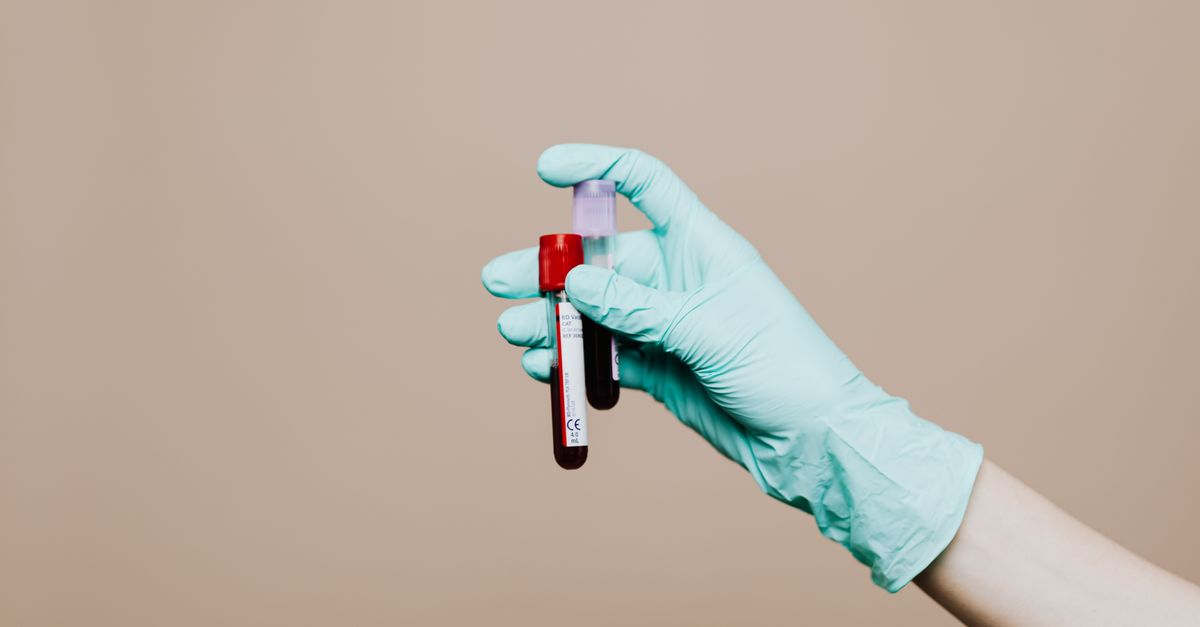
What Happens to FSH Levels in the Postpartum Period?
FSH (follicle-stimulating hormone) remains suppressed for weeks to several months after delivery, driven primarily by elevated prolactin from breastfeeding or by residual GnRH suppression if not nursing. For most postpartum women this is a normal, temporary state. The exception: if FSH stays blunted well beyond 6 months postpartum — especially after weaning — it warrants evaluation for secondary hypothalamic dysfunction, thyroid disruption, or pituitary pathology.
---
Why FSH Drops After Delivery
During pregnancy, the hypothalamic-pituitary-ovarian (HPO) axis is systematically shut down by high placental estrogen, progesterone, and hCG. At delivery, placental hormones clear rapidly, but the HPO axis does not simply switch back on. Two distinct suppressive forces take over:
Hyperprolactinemia. Breastfeeding drives prolactin into the 100–300 ng/mL range — levels that tonically inhibit GnRH pulsatility. Without normal GnRH pulses, the pituitary cannot release meaningful quantities of FSH or LH (Tay et al., Obstetrics & Gynecology 1996; PMID: 8765268).
Residual hypothalamic suppression. Even in non-breastfeeding mothers, it can take 4–8 weeks for GnRH pulse amplitude and frequency to normalize. Studies using frequent blood sampling have confirmed that LH pulse frequency is markedly blunted in the first weeks postpartum regardless of feeding status (McNeilly et al., Journal of Clinical Endocrinology & Metabolism 1992; PMID: 1639942).
The clinical upshot: FSH measured in the first 4–6 weeks postpartum will often look falsely low — sometimes in the 1–3 IU/L range, which overlaps with the suppressed values seen in hypothalamic amenorrhea. This does not indicate a problem; it reflects the expected hormonal reset.
The Timeline of FSH Recovery
Recovery follows a fairly predictable pattern when breastfeeding is the dominant variable:
| Postpartum Phase | Typical FSH Range | Notes |
|---|---|---|
| 0–6 weeks | 1–4 IU/L | Prolactin dominant; GnRH pulses sparse |
| 6–12 weeks (non-nursing) | 3–8 IU/L | HPO axis beginning to recover |
| 3–6 months (exclusive nursing) | 1–5 IU/L | Lactational amenorrhea maintained |
| After weaning | 4–10 IU/L within 4–8 weeks | First ovulation typically precedes first period |
| 6–12 months post-weaning | Normal follicular-phase range (3–10 IU/L) | Full axis recovery expected |
Women who are not breastfeeding typically see their first ovulation around 4–6 weeks postpartum, with FSH rising to follicular-phase levels shortly before that event. A landmark analysis of daily hormone profiles confirmed that the first postpartum LH surge — and subsequent FSH rise — occurred as early as 27 days in non-lactating women (Glasier et al., Clinical Endocrinology 1983; PMID: 6573416).
---
How Prolactin Suppresses FSH: The Mechanism in Detail
Understanding why FSH stays low helps you interpret labs more accurately and avoid unnecessary interventions.
Prolactin acts at multiple levels of the reproductive axis. At the hypothalamus, it suppresses kisspeptin neuron firing. Kisspeptin neurons in the arcuate nucleus are the master pacemakers of GnRH pulsatility — without their regular activity, GnRH is not secreted in the rhythmic bursts the pituitary needs to produce LH and FSH. This is why lactational amenorrhea is so effective as a contraceptive method: it disrupts the chain of signaling at the very first step.
At the pituitary level, sustained high prolactin also downregulates GnRH receptor sensitivity, meaning that even if some GnRH pulses occur, the gonadotroph cells respond less robustly. The net effect is a disproportionately low FSH-to-LH ratio that can persist for weeks after weaning begins, because it takes time for both kisspeptin tone and pituitary receptor density to normalize.
Succkling frequency matters enormously. Studies tracking nursing behavior alongside hormone profiles found that women nursing more than 6 times per day maintained significantly higher prolactin and significantly lower FSH than women nursing 3–4 times per day (Tay et al., Obstetrics & Gynecology 1996; PMID: 8765268). This dose-response relationship is why partial weaning — dropping one or two feeds — often triggers a faster FSH rebound than many clinicians expect.
---
When Postpartum Thyroid Disruption Mimics Low FSH Problems
Postpartum thyroiditis affects an estimated 5–10% of women in the first year after delivery. It matters for FSH because thyroid dysfunction — both hypo- and hyperthyroid phases — independently disrupts GnRH pulsatility and gonadotropin secretion. A woman with subclinical hypothyroidism (elevated TSH, low-normal free T4) can have a blunted FSH response to GnRH stimulation even months after weaning, making it appear that the HPO axis has not recovered when the real problem is inadequate thyroid hormone signaling.
For a detailed look at how TSH shifts in the months after delivery, see what happens to TSH levels in the postpartum period. Similarly, what happens to vitamin D during postpartum is relevant here because vitamin D receptors are expressed on GnRH neurons and vitamin D deficiency — very common in the postpartum period — can impair GnRH pulsatility independently of prolactin levels.
The practical implication: before attributing persistent FSH suppression to pituitary pathology, clinicians should check TSH, free T4, and 25-OH vitamin D as part of the workup. In many cases, correcting subclinical hypothyroidism or repleting vitamin D is sufficient to restore normal gonadotropin secretion without any further intervention.
---
How Stress Affects FSH Recovery Postpartum
Stress is a well-documented suppressor of the HPO axis at the hypothalamic level, and the postpartum period is among the most psychologically and physiologically demanding of a woman's life. Cortisol and CRH (corticotropin-releasing hormone) both inhibit GnRH neuron firing. In the postpartum context, this means that a new mother managing sleep deprivation, nutritional depletion, and psychological stress is operating with a cortisol environment that actively works against HPO axis recovery.
Research on hypothalamic amenorrhea in athletes and women with caloric restriction provides a useful model. In those populations, FSH and LH suppression is tightly correlated with 24-hour cortisol output and inversely correlated with caloric adequacy (Loucks et al., Journal of Clinical Endocrinology & Metabolism 2003; PMID: 12519869). The postpartum period stacks multiple of these same suppressors simultaneously — poor sleep, energy deficit from breastfeeding caloric demands, and emotional stress — which is why some women's FSH remains depressed well past the expected recovery window.
Adaptogenic support for the HPA axis is not a direct FSH therapy, but normalizing cortisol burden removes one of the primary brakes on GnRH pulsatility. Ashwagandha root extract (KSM-66) at 600 mg/day reduced serum cortisol by 27.9% and improved subjective stress scores significantly over 60 days in a randomized controlled trial of chronically stressed adults (Chandrasekhar et al., Indian Journal of Psychological Medicine 2012; PMID: 23439798). Reducing HPA axis hyperactivity in the postpartum period may help create the hormonal environment the HPO axis needs to resume normal FSH secretion.
---
Supplements for FSH: What the Evidence Actually Supports
The supplement category labeled "supplements for FSH" is broad and sometimes misleading. Most products marketed to "boost FSH" are actually targeting underlying drivers of suppression rather than directly stimulating FSH secretion. Understanding this distinction is important.
Myo-Inositol
Myo-inositol (2–4 g/day) is the most studied supplement for FSH normalization, though the majority of trials focus on women with polycystic ovary syndrome (PCOS) rather than postpartum populations specifically. Myo-inositol improves insulin signaling in granulosa cells and appears to support more regular FSH pulsatility, with trials showing improved follicular development and normalized FSH-to-LH ratios in PCOS (Unfer et al., Gynecological Endocrinology 2012; PMID: 22296283). Whether this translates to postpartum HPO axis recovery is mechanistically plausible but not directly studied.
Vitamin D3
Vitamin D is not a gonadotropin secretagogue in the strict sense, but vitamin D deficiency is associated with blunted GnRH sensitivity and suboptimal FSH levels across multiple populations. Supplementation at 2,000–4,000 IU/day in deficient women has been shown to improve menstrual regularity — a downstream proxy for normalized FSH secretion — particularly in women with baseline 25-OH D levels below 20 ng/mL. Given that postpartum vitamin D deficiency is extremely prevalent, this is one of the highest-yield interventions available.
Omega-3 Fatty Acids (EPA/DHA)
Omega-3s support prostaglandin metabolism and reduce systemic inflammation — both relevant to hypothalamic signaling. In women with dysmenorrhea and irregular cycles, omega-3 supplementation (1,800–2,700 mg/day combined EPA + DHA) was associated with improvements in cycle regularity, suggesting a modulatory effect on the HPO axis environment that may be relevant during postpartum recovery.
---
Supplements to Lower FSH: A Different Clinical Context
The goal of lowering FSH applies in a completely different clinical scenario: women with elevated FSH, which typically signals diminished ovarian reserve (DOR) or early perimenopause. This is not the typical postpartum picture — in fact, it is the opposite problem. But it is worth addressing because some postpartum women do present with unexpectedly elevated FSH after weaning, which can indicate that ovarian function was compromised prior to or during pregnancy.
In women with elevated FSH and DOR, the evidence-based supplement protocol looks quite different:
- CoQ10 (Ubiquinol, 400–600 mg/day): Mitochondrial support for oocyte energy metabolism. A randomized trial in women with poor ovarian response undergoing IVF showed that ubiquinol at 600 mg/day improved oocyte quality markers compared to placebo (Xu et al., Journal of Ovarian Research 2018; PMID: 30053853).
- DHEA (25–75 mg/day): Used in fertility medicine to improve ovarian response in DOR; can reduce elevated FSH in some patients over a 3–6 month course. Should only be used under clinical supervision given androgenic effects.
- Melatonin (3 mg/day): Antioxidant role in follicular fluid; associated with improved oocyte quality in IVF studies. Not directly related to FSH levels but addresses the oxidative environment that damages follicles.
For women dealing with elevated FSH and cardiovascular risk markers simultaneously — which can coexist in perimenopause — understanding the full lipid picture is important. Resources like what supplements lower ApoB and supplements that lower triglycerides provide evidence-based guidance for that concurrent clinical picture.
---
What This Means for Your Formula
Ones doesn't offer a single "FSH supplement" because FSH normalization is almost always about addressing upstream drivers rather than targeting the hormone directly. Based on the mechanisms reviewed above, the most relevant Ones ingredients for postpartum women with delayed HPO axis recovery are:
Vitamin D3 + K2 (MK-7): Ones includes vitamin D3 paired with MK-7 to support absorption and cardiovascular safety at doses calibrated to your actual 25-OH D level from blood work — not a one-size-fits-all 1,000 IU. For postpartum women where vitamin D insufficiency is contributing to GnRH suppression, getting to a replete 25-OH D level (50–70 ng/mL) removes a meaningful brake on HPO axis recovery.
KSM-66 Ashwagandha (600 mg): Where lab data or wearable stress markers suggest elevated HPA axis activity, Ones includes KSM-66 at the full 600 mg clinical dose — the same dose used in the Chandrasekhar cortisol-reduction trial. Normalizing cortisol burden in the postpartum period creates a more permissive hormonal environment for GnRH pulsatility to resume.
Omega-3 (EPA/DHA): Ones formulas incorporate pharmaceutical-grade omega-3 at doses matched to your inflammatory markers, supporting the prostaglandin environment and reducing systemic inflammation that can blunt hypothalamic sensitivity.
For postpartum women whose labs also reveal thyroid disruption — a common co-finding — the T4 to T3 conversion supplements article explains how supporting thyroid hormone activation complements HPO axis recovery. And because ferritin levels postpartum are frequently depleted, iron repletion is often a parallel priority that Ones addresses when blood work confirms low stores.
---
Key Takeaways
- FSH suppression in the first 3–6 months postpartum is physiologically normal, driven primarily by hyperprolactinemia from breastfeeding and residual GnRH axis suppression.
- Non-breastfeeding women recover faster — first ovulation can occur as early as 27 days postpartum, with FSH rising to follicular-phase levels within 4–8 weeks.
- Suckling frequency drives prolactin, which drives FSH suppression — even partial weaning can trigger measurable FSH rebound within weeks.
- Thyroid dysfunction and vitamin D deficiency are common postpartum co-factors that can extend FSH suppression beyond the expected window and should be evaluated before assuming pituitary pathology.
- Chronic stress and cortisol burden actively suppress GnRH pulsatility, making HPA axis support a legitimate adjunct strategy for delayed FSH recovery — ashwagandha at 600 mg/day has the strongest cortisol-reduction evidence in this context.
- Elevated FSH postpartum is a different problem from suppressed FSH and may indicate diminished ovarian reserve that predated pregnancy; CoQ10 (ubiquinol) and targeted antioxidant support are the most evidence-based interventions in that scenario.
- Always consult a healthcare provider before interpreting postpartum hormone panels in isolation — FSH values only make clinical sense alongside prolactin, LH, TSH, and estradiol in context.