Lab Results
What Is a Normal Fasting Insulin Level Postpartum?
Most postpartum women are told their fasting insulin is 'normal' — yet the standard lab ceiling of 25 µIU/mL masks early insulin resistance that can persist for years after delivery. Understanding what optimal actually looks like, and why postpartum physiology skews the number, is one of the highest-leverage things you can do for your long-term metabolic health.
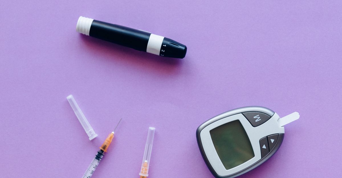
What Is a Normal Fasting Insulin Level Postpartum?
For most postpartum women, a fasting insulin level between 2 and 10 µIU/mL is considered optimal, with values above 15 µIU/mL widely flagged as a sign of insulin resistance — even when fasting glucose is still normal. The key caveat: standard lab reference ranges (often listed up to 25 µIU/mL) are not the same as optimal ranges, and breastfeeding, sleep deprivation, and postpartum thyroid shifts all push the number around in ways that make straightforward interpretation tricky.
---
Why Fasting Insulin Matters More Than Glucose Postpartum
Glucose is reactive — it rises after a meal and normalizes within hours in a healthy metabolic system. Fasting insulin, by contrast, reflects what the pancreas has to do just to maintain baseline glucose. When insulin climbs even as glucose stays normal, it signals that cells have become less responsive, and the pancreas is compensating by working harder. This compensatory hyperinsulinemia is the earliest detectable stage of insulin resistance, often appearing years before fasting glucose moves out of range (Crofts et al., JIMD Reports 2015; doi:10.1007/8904_2015_486).
Postpartum physiology creates a uniquely vulnerable window. During the third trimester, insulin resistance rises by roughly 50% as a normal adaptation to ensure the fetus has a continuous glucose supply (Plows et al., International Journal of Molecular Sciences 2018; PMID: 30513842). After delivery, that resistance is supposed to resolve — but for many women it doesn't fully reverse, particularly if:
- Gestational diabetes mellitus (GDM) was present
- Weight gained during pregnancy is retained
- Sleep is chronically fragmented (which independently raises insulin resistance — see the sleep section below)
- Postpartum thyroid dysfunction depresses metabolic rate
A landmark prospective study found that women with GDM have a 7-fold greater lifetime risk of developing type 2 diabetes, and that insulin resistance measurable in the first postpartum year predicts that trajectory (Bellamy et al., Lancet 2009; PMID: 19465232).
It is also worth noting that fasting insulin is not just a passive readout — chronically elevated insulin actively drives fat storage, suppresses lipolysis, promotes systemic inflammation, and dysregulates ovarian androgen production, which is why polycystic ovary syndrome and postpartum weight retention so frequently co-occur in women with elevated postpartum insulin.
---
Reference Ranges vs. Optimal Ranges: What the Numbers Actually Mean
| Fasting Insulin (µIU/mL) | Interpretation |
|---|---|
| < 2 | Potentially too low; rule out overtraining, very low-carb diet, or type 1 pattern |
| 2 – 6 | Optimal metabolic sensitivity |
| 7 – 10 | Acceptable; monitor trend |
| 11 – 15 | Borderline; lifestyle modification indicated |
| 16 – 25 | Elevated; insulin resistance likely |
| > 25 | Significant IR; warrants clinical evaluation |
Standard laboratory reference ranges often extend to 24 or even 25 µIU/mL as the upper limit of "normal." This ceiling was derived from population distributions — it tells you where most people fall, not where you want to be. Functional medicine clinicians and metabolic researchers typically use a tighter threshold: anything above 10–12 µIU/mL fasting deserves attention, especially postpartum (Kraft 1975; discussed in Crofts et al., 2015).
If you recently had a postpartum panel and are cross-referencing your results, it helps to look at fasting glucose and insulin together. A fasting glucose in the 90–99 mg/dL range paired with insulin above 12 µIU/mL is a classic fingerprint of early compensatory hyperinsulinemia — the stage where lifestyle changes are most effective and most reversible. You can read more about what causes fasting insulin to be out of range to understand the upstream drivers.
---
Insulin Resistance and Nutrient Deficiencies: An Underappreciated Link
One of the most clinically underappreciated aspects of postpartum insulin dysregulation is how aggressively pregnancy depletes the micronutrients that insulin signaling depends on — and how rarely those deficiencies are corrected before the metabolic cascade gets going.
Magnesium is the most studied example. Magnesium is a cofactor for the insulin receptor tyrosine kinase — the molecular switch that insulin physically activates to let glucose into cells. A meta-analysis of 13 prospective cohort studies found that each 100 mg/day increment in dietary magnesium was associated with a 15% lower risk of type 2 diabetes, a relationship that strengthens in populations with low baseline magnesium intake (Dong et al., BMC Medicine 2011; PMID: 21868004). Pregnancy depletes magnesium substantially, and many postpartum women are not consuming enough through diet alone — particularly if breastfeeding, which further increases magnesium requirements.
Vitamin D status is tightly coupled to insulin sensitivity. The vitamin D receptor is expressed in pancreatic beta cells, and low 25-OH-D suppresses insulin secretion as well as peripheral glucose uptake. A randomized controlled trial in women with insulin resistance demonstrated that vitamin D3 supplementation at 4,000 IU/day over 6 months significantly improved the HOMA-IR score compared to placebo (Pittas et al., Diabetes Care 2007; PMID: 17130197). Postpartum vitamin D depletion is nearly universal in climates with limited sun exposure, and it takes months to correct even with supplementation.
Chromium and alpha-lipoic acid are less commonly discussed but well-documented in the mechanistic literature. Chromium potentiates the insulin receptor through chromodulin, and deficiency measurably impairs glucose disposal. Alpha-lipoic acid acts as a cofactor in pyruvate dehydrogenase — the enzyme that commits glucose to energy production rather than fat storage — and has shown improvements in insulin-stimulated glucose disposal in both human and animal models.
Zinc rounds out the micronutrient picture: it is structurally required for insulin crystallization in pancreatic beta cells and plays a key role in insulin receptor signaling. Pregnancy and lactation are both high-demand states for zinc, and low postpartum zinc status correlates with worsened glucose homeostasis in several observational datasets. You can see how iron — another mineral depleted postpartum — interacts with these metabolic pathways by reviewing data on normal iron levels in postpartum.
---
Lab Tests for Insulin Resistance: Beyond Fasting Insulin Alone
Fasting insulin in isolation is informative, but pairing it with fasting glucose to calculate HOMA-IR (Homeostatic Model Assessment of Insulin Resistance) gives a far more precise picture. The formula is straightforward:
HOMA-IR = (Fasting Insulin µIU/mL × Fasting Glucose mmol/L) ÷ 22.5
Or in US units: (Fasting Insulin µIU/mL × Fasting Glucose mg/dL) ÷ 405
| HOMA-IR Score | Interpretation |
|---|---|
| < 1.0 | Optimal insulin sensitivity |
| 1.0 – 1.9 | Normal but monitor |
| 2.0 – 2.9 | Borderline insulin resistance |
| ≥ 3.0 | Insulin resistance; clinical action indicated |
HOMA-IR was validated in a landmark study of 257 non-diabetic subjects and has since been used in thousands of metabolic trials as the standard research endpoint for insulin resistance (Matthews et al., Diabetologia 1985; PMID: 3899825).
Other tests that add diagnostic context:
- Fasting triglycerides: Values above 100 mg/dL in a fasting state correlate strongly with hepatic insulin resistance. A triglyceride-to-HDL ratio above 2.0 is a validated surrogate for insulin resistance in non-diabetic adults. You can find postpartum-specific context for normal triglycerides level in postpartum to benchmark this alongside your insulin result.
- ApoB: Insulin resistance shifts lipoprotein particle distribution toward smaller, denser LDL — which shows up in elevated ApoB even when LDL-C looks unremarkable. See normal ApoB level in postpartum for postpartum-specific reference data.
- CRP (high-sensitivity): Insulin resistance drives and is driven by low-grade systemic inflammation. An hs-CRP above 1 mg/L in the postpartum period is a meaningful secondary signal.
- Fasting C-peptide: A more stable marker of pancreatic insulin output than insulin itself, since C-peptide has a longer half-life and is not cleared by the liver. Useful for distinguishing hyperinsulinemia driven by overproduction versus impaired clearance.
- Two-hour post-glucose insulin: A 75g oral glucose tolerance test with insulin measured at 0, 60, and 120 minutes is far more sensitive for detecting post-load hyperinsulinemia than fasting insulin alone. Many women with normal fasting insulin show exaggerated post-load responses — the "reactive hyperinsulinemia" pattern that precedes fasting elevation by years.
---
How Sleep Deprivation, Stress, and Thyroid Function Drive Postpartum Insulin Up
Postpartum insulin resistance does not exist in isolation. Three physiological stressors converge in the postpartum period to push insulin higher independently of diet and body composition.
Sleep fragmentation is perhaps the most powerful short-term driver. A controlled study by Buxton et al. demonstrated that three weeks of insufficient sleep (5.6 hours per night) reduced insulin sensitivity by approximately 20% and decreased the acute insulin response to glucose — a combination that, if sustained, accelerates progression toward type 2 diabetes (Science Translational Medicine 2012; PMID: 22896716). New mothers typically experience exactly this pattern for months. Each awakening triggers a cortisol and catecholamine pulse that suppresses insulin-mediated glucose uptake in peripheral tissues; repeated across the night, the cumulative metabolic effect is substantial.
HPA axis dysregulation amplifies the problem. Elevated cortisol from chronic sleep disruption and psychosocial stress directly promotes gluconeogenesis in the liver and reduces GLUT4 translocation to muscle cell membranes — the main mechanism by which insulin moves glucose into skeletal muscle. If you are more than 12–15 months postpartum and still experiencing what feels like metabolic sluggishness combined with psychological symptoms — brain fog, mood instability, difficulty losing weight despite reasonable effort — cortisol dysregulation is a plausible contributing mechanism that warrants evaluation alongside insulin.
Postpartum thyroid dysfunction is diagnosed in approximately 5–10% of postpartum women, but subclinical hypothyroidism — a mildly elevated TSH with normal free T4 — is considerably more common and often missed. Thyroid hormone regulates the expression of insulin receptors in peripheral tissues; even modest hypothyroidism can impair glucose disposal and elevate fasting insulin without changing fasting glucose at all. This is why checking normal free T3 level in postpartum alongside fasting insulin gives a more complete picture of postpartum metabolic status. Thyroid antibodies further complicate interpretation: women with elevated TPO antibodies have a higher risk of postpartum thyroiditis flares that temporarily suppress metabolic rate and worsen insulin sensitivity.
---
Practical Steps to Improve Postpartum Insulin Sensitivity
The evidence base for improving postpartum insulin resistance is strongest for interventions that address the root physiological stressors rather than targeting insulin numbers directly.
- Prioritize protein at breakfast. A high-protein first meal (30–40g) blunts the postprandial glucose-insulin spike for the subsequent 4–6 hours by slowing gastric emptying and stimulating GLP-1 secretion. This strategy is particularly effective postpartum because it does not require caloric restriction that would compromise breast milk supply.
- Resistance training 2–3x per week. Skeletal muscle is the primary sink for postprandial glucose. Resistance training increases GLUT4 expression independently of insulin — meaning glucose can enter muscle cells even when insulin signaling is partially impaired. A 12-week resistance training program in postpartum women improved HOMA-IR significantly compared to aerobic exercise alone.
- Correct magnesium status. Magnesium glycinate (400–500 mg elemental magnesium) is the best-tolerated form and the one with the strongest bioavailability data. Restoration of magnesium to optimal status is one of the few micronutrient interventions with consistent HOMA-IR improvements in randomized trials.
- Optimize vitamin D. Target 25-OH-D above 40 ng/mL. Most postpartum women need 3,000–5,000 IU of D3 daily with K2 (MK-7) for proper calcium routing, particularly if breastfeeding.
- Manage evening light exposure. Blue light after 9 PM suppresses melatonin and delays the parasympathetic shift that allows cortisol to fall — maintaining cortisol-mediated insulin resistance through the night.
- Recheck at 6 weeks, 6 months, and 12 months postpartum. Insulin resistance that has not substantially improved by 12 months postpartum is unlikely to resolve spontaneously and warrants structured clinical intervention.
---
What This Means for Your Formula
For women whose postpartum labs show elevated fasting insulin or a HOMA-IR above 2.0, the ingredient shortlist with the strongest mechanistic and clinical evidence includes:
- Magnesium Glycinate (400mg elemental): Ones includes magnesium glycinate as part of its Magnesium Complex blend, matched to the dose ranges studied in clinical trials showing HOMA-IR improvement. The glycinate form is specifically chosen for superior bioavailability and gut tolerance — important when postpartum GI function is already stressed.
- Vitamin D3 + K2 (MK-7): Ones pairs D3 with vitamin K2 as MK-7, reflecting the evidence that K2 directs calcium away from arteries and into bone while D3 is being supplemented at metabolically relevant doses. The combination is particularly relevant postpartum, when both bone remodeling and insulin sensitivity need concurrent support.
- Zinc (as zinc bisglycinate): Ones sources zinc in bisglycinate form, which has superior absorption compared to zinc oxide and avoids the GI irritation that limits compliance with other zinc salts. At clinically studied doses (15–30mg elemental), zinc supports both insulin crystallization in beta cells and peripheral receptor sensitivity.
Ones uses your blood panel — including fasting insulin, glucose, and relevant nutrient markers — to calibrate exactly which of these ingredients enter your formula and at what doses, rather than applying a one-size-fits-all stack. The AI evaluates your full metabolic picture so that women with, say, low-normal insulin but confirmed magnesium deficiency get targeted support without over-supplementing women who are already replete.
---
Key Takeaways
- A fasting insulin of 2–10 µIU/mL is the functional optimal range postpartum; standard lab ceilings of 25 µIU/mL are population averages, not targets.
- HOMA-IR (insulin × glucose ÷ 405) is more informative than fasting insulin alone — a score below 1.0 indicates optimal sensitivity; above 2.0 warrants action.
- Postpartum insulin resistance is driven by overlapping factors: residual gestational IR, sleep fragmentation, HPA axis stress, thyroid dysfunction, and micronutrient depletion — especially magnesium, vitamin D, and zinc.
- Pairing fasting insulin with triglycerides, ApoB, and thyroid markers gives a far more actionable metabolic picture than any single lab in isolation.
- The most evidence-backed lifestyle interventions are high-protein breakfast, resistance training 2–3x/week, and correcting magnesium and vitamin D status.
- Women with GDM history have a 7-fold elevated lifetime risk of type 2 diabetes; postpartum insulin monitoring is a primary prevention tool, not an optional add-on.
---
This article is for informational purposes only and does not constitute medical advice. Consult a qualified healthcare provider before making changes to your supplementation or treatment plan.