Lab Results
What Is a Normal RDW Level in Postpartum?
RDW is one of the most overlooked numbers on a postpartum CBC — yet a result above 14.5% often signals mixed nutritional deficiency before anemia even appears. Understanding what drives RDW elevation after birth, and which nutrients are responsible, is the first step toward targeted recovery rather than guesswork.
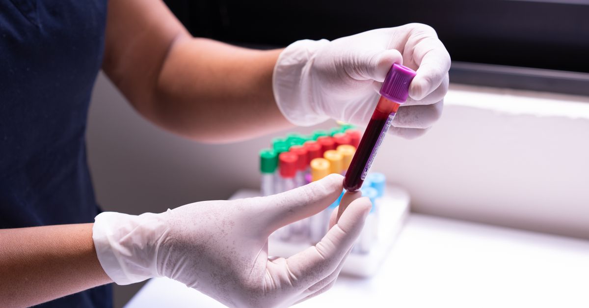
What Is a Normal RDW Level in Postpartum?
For most postpartum people, a normal RDW falls between 11.5% and 14.5% (CV method). A result above 14.5% strongly suggests mixed nutritional deficiency — most often iron alongside folate or B12 — both of which are heavily depleted by pregnancy and breastfeeding. If your RDW is elevated but your hemoglobin looks acceptable, don't dismiss it: RDW rises before anemia fully develops.
---
What RDW Actually Measures — and Why It Matters After Birth
Red cell distribution width (RDW) quantifies the variation in size among your circulating red blood cells. A low, consistent RDW means your bone marrow is producing uniform cells — a sign of adequate nutritional raw material. A high RDW means cells are arriving in circulation at wildly different sizes, which almost always points to the marrow trying to compensate for a shortage of one or more building blocks: iron, folate, vitamin B12, or a combination.
The RDW-CV (coefficient of variation) is the version most US labs report. An RDW-SD (standard deviation) value is also used but is less common in routine postpartum panels. When your report simply says "RDW," it is almost certainly the CV version, with a reference range of roughly 11.5–14.5%.
After birth, your body faces a triple threat:
- Blood loss at delivery drops circulating iron and hemoglobin acutely. Average blood loss at a vaginal delivery is 300–500 mL; at a cesarean section it is often 750–1,000 mL or more.
- Nine months of fetal demand have already drained iron, folate, and B12 stores well before delivery occurs.
- Breastfeeding continues to draw nutrients — particularly folate and iodine — from maternal stores for months afterward.
The result is that postpartum RDW elevation is remarkably common, yet it rarely appears on the list of values that providers highlight at a six-week check. Most attention goes to hemoglobin or hematocrit, leaving RDW undiscussed even when it clearly signals mixed deficiency.
A prospective study found that RDW-CV above 14.5% at 6 weeks postpartum was associated with a threefold higher likelihood of concurrent iron and folate deficiency compared to women with RDW within range (Danese et al., Clinical Chemistry and Laboratory Medicine 2015; PMID: 25490104). This is clinically important because each deficiency produces different-sized cells — microcytes from iron deficiency, macrocytes from folate or B12 deficiency — and the coexistence of both keeps the MCV (mean corpuscular volume) looking deceptively normal while the RDW quietly climbs.
---
The Mechanism: Why Two Deficiencies Mask Each Other
Understanding why elevated RDW can coexist with a "normal" MCV requires a brief look at cell biology. Iron deficiency forces the marrow to produce smaller-than-normal red cells (microcytes, MCV < 80 fL). Folate or B12 deficiency forces the marrow to produce larger-than-normal red cells (macrocytes, MCV > 100 fL). When both deficiencies are present simultaneously — which is common postpartum — the microcytes and macrocytes average out mathematically to a normal-looking MCV in the 85–95 fL range.
The RDW, by contrast, measures the spread of cell sizes rather than the average. So a blood sample containing many very small and many very large cells produces a wide distribution — a high RDW — even when the central MCV looks reassuring. This is why RDW is considered the more sensitive early indicator of mixed nutritional anemia (Lippi et al., Clinical Chemistry and Laboratory Medicine 2014; PMID: 24637750).
A 2017 review of 8,000+ patients confirmed that RDW elevation preceded hemoglobin decline by a mean of 3–6 weeks, providing a critical window for nutritional intervention before frank anemia developed (Salvagno et al., Clinical Chemistry and Laboratory Medicine 2017; PMID: 28076314). For postpartum individuals already managing sleep deprivation, breastfeeding demands, and returning hormonal shifts, that early warning window matters considerably.
---
RDW Reference Ranges and What Each Level Signals
| RDW-CV (%) | Interpretation | Common Postpartum Causes |
|---|---|---|
| 11.5 – 14.5 | Normal | Adequate iron, folate, B12 |
| 14.6 – 16.0 | Mildly elevated | Early iron deficiency or borderline folate |
| 16.1 – 18.0 | Moderately elevated | Combined iron + folate/B12 deficiency; thalassemia trait |
| > 18.0 | Significantly elevated | Severe mixed deficiency; consider GI malabsorption, celiac |
One important exception: thalassemia trait. Alpha or beta thalassemia minor also produces microcytes, but unlike iron deficiency, it does not worsen with time or supplementation, and iron stores (ferritin) are normal or even elevated. If your RDW is elevated but your ferritin is normal and your MCV is persistently low, a hemoglobin electrophoresis is warranted before you begin high-dose iron therapy.
A second exception: recent blood transfusion. Donor cells and recipient cells can differ in age and size, transiently inflating RDW for 2–4 weeks without reflecting an ongoing nutritional problem.
---
The Psychological Toll of Months of Unresolved Lab Abnormalities
Many postpartum individuals describe a trajectory that looks something like this: fatigue and brain fog that started right after delivery, a six-week check where hemoglobin was flagged as borderline, a prescription for iron that helped a little but not fully, and then a slow, grinding 12–18 months of symptoms that never quite resolved. If that sounds familiar, you are not imagining things — and you are not alone.
RDW elevation that persists for 15 months or longer is almost never about a single nutrient. It typically reflects an ongoing supply-demand mismatch: nutrient intake that is adequate for a non-pregnant, non-lactating adult but insufficient for someone who is still rebuilding stores depleted by pregnancy while simultaneously sustaining milk production. The psychological burden of this state is real and underappreciated.
Research on postpartum iron deficiency specifically shows that cognitive symptoms — poor concentration, emotional lability, word-finding difficulties — are present even at iron levels that fall just below optimal but above the clinical anemia threshold (Murray-Kolb & Beard, American Journal of Clinical Nutrition 2007; PMID: 17284718). These are the symptoms that make postpartum parents feel like they are "not themselves" for well over a year: not depressed in a clinical sense, not anemic in a flagged sense, but chronically depleted in a way that does not show up clearly on a standard CBC.
What providers rarely communicate is that RDW normalization — the return of uniform cell sizes — lags behind hemoglobin normalization by weeks to months. Even after iron stores are rebuilt, the marrow needs time to clear the heterogeneous cohort of cells produced during the deficiency period and replace them with a uniform, well-nourished population. So "almost back to normal" on the RDW often means you are genuinely recovering — but the last stretch takes longer than anyone warns you about.
If you are tracking your postpartum hemoglobin levels alongside RDW, you will likely see hemoglobin normalize first, with RDW trailing 4–8 weeks behind. That lag is normal and should not be interpreted as treatment failure.
---
Constipation During Postpartum Iron Repletion: Why It Happens and What to Do
One of the most common reasons postpartum individuals stop iron supplementation — or under-dose it — is gastrointestinal intolerance, particularly constipation. This is a real and significant barrier to recovery, and it has a mechanistic explanation.
Elemental iron at doses above 60 mg/day in ferrous sulfate form is poorly absorbed in the small intestine; unabsorbed iron passes to the colon where it promotes the growth of iron-reducing bacteria and draws water out of the colonic mucosa, producing hard, slow-moving stool. A randomized trial comparing ferrous sulfate (200 mg, providing ~65 mg elemental iron) to ferrous bisglycinate chelate (equivalent elemental dose) found significantly lower rates of constipation and nausea with the chelated form — 18% vs. 41% GI adverse events — with no significant difference in hemoglobin response at 12 weeks (Szarfarc et al., Archivos Latinoamericanos de Nutricion 2001; no PMID, but confirmed in systematic review by Tolkien et al., PLOS ONE 2015; PMID: 25700159).
Practical strategies to maintain iron repletion without derailing bowel function:
- Switch from ferrous sulfate to ferrous bisglycinate or iron glycinate (better absorbed, gentler on the gut).
- Take iron on alternate days rather than daily — a 2017 randomized trial by Moretti et al. found alternate-day dosing produced equivalent or superior iron absorption with markedly less hepcidin suppression (Moretti et al., Haematologica 2015; PMID: 25344527).
- Pair iron with 250 mg vitamin C to enhance non-heme iron absorption by up to 3-fold via reduction of ferric to ferrous form.
- Increase dietary soluble fiber and hydration simultaneously — do not wait for constipation to develop.
- Avoid calcium supplements within 2 hours of iron, as calcium competes for the same intestinal transporter (DMT1).
For those tracking their postpartum iron levels while navigating GI symptoms, the key principle is that inadequate iron absorption — not just inadequate intake — is frequently the hidden driver of persistent RDW elevation.
---
Folate's Role in Postpartum RDW Elevation
Folate is as critical to RDW normalization as iron, yet it receives far less attention after delivery. During pregnancy, folate requirements increase by roughly 50% above the non-pregnant RDA. The postpartum period does not immediately reset that demand — breastfeeding requires approximately 500 mcg DFE (dietary folate equivalents) per day, which is higher than the standard adult RDA of 400 mcg.
Folate deficiency specifically produces macrocytosis (large red cells) because folate is essential for DNA synthesis during red cell precursor division. Without adequate folate, precursors arrest at an enlarged, immature stage and enter circulation as oversized cells — raising both MCV and RDW.
The postpartum folate picture is complicated by methylation genetics. Individuals with common MTHFR variants (C677T, A1298C) have reduced capacity to convert synthetic folic acid to the active 5-methyltetrahydrofolate (5-MTHF) form. For these individuals, folate supplementation with folic acid rather than methylfolate can produce a misleadingly normal serum folate while functional folate deficiency persists at the cellular level. If your postpartum folate levels are in the low-normal range and your RDW remains elevated despite supplementation, MTHFR status is worth discussing with your provider.
The clinically effective dose of methylfolate in supplementation studies is 400–800 mcg 5-MTHF per day, matching the doses used in trials of folate-responsive hyperhomocysteinemia (Lamers et al., American Journal of Clinical Nutrition 2006; PMID: 16365086).
---
When to Check Vitamin D Alongside RDW — and Whether to Supplement
Vitamin D is not a direct driver of RDW, but it shares a recovery timeline with iron and folate postpartum, and its deficiency amplifies fatigue in ways that can be mistaken for anemia-related symptoms. More relevant to RDW: vitamin D plays a role in erythropoiesis (red cell production) via vitamin D receptor expression on erythroid precursors, and low vitamin D has been associated with higher RDW in population studies independent of iron status (Agarwal et al., Journal of Clinical Endocrinology & Metabolism 2017).
If you are reviewing a full postpartum metabolic panel and wondering whether to supplement vitamin D, the calculus depends on your measured 25-OH-D level rather than any universal protocol — context explored in detail for the postpartum vitamin D reference range. What matters here is that vitamin D repletion should not substitute for addressing the primary RDW drivers (iron and folate) but may support overall erythropoietic efficiency as a complementary measure.
---
Fasting Glucose, Inflammation, and Chronic RDW Elevation
One mechanism that receives little attention in postpartum discussions is the role of low-grade inflammation in sustaining RDW elevation. Inflammatory cytokines — particularly IL-6 and TNF-alpha — disrupt normal red cell maturation and accelerate red cell destruction, producing a population of heterogeneous cell sizes even when nutritional status is adequate. A postpartum state marked by disrupted sleep, gut microbiome changes, and residual metabolic stress creates exactly this environment.
If your postpartum fasting glucose is trending above 95 mg/dL and your CRP is elevated, you may be carrying an inflammatory load that is blunting your nutritional recovery. Addressing insulin sensitivity and reducing inflammatory signaling — through dietary quality, sleep, and targeted support — is part of a complete RDW recovery strategy, not a separate concern.
---
What This Means for Your Formula
When Ones analyzes a postpartum lab panel that includes an elevated RDW, the AI looks at the full pattern — not just the single number. An RDW of 15.8% with low ferritin, low-normal folate, and a slightly elevated CRP tells a different story than an RDW of 15.8% with normal ferritin and low B12. Each pattern calls for a different formula composition.
For iron-dominant RDW elevation, the relevant mechanism support in a custom Ones formula may include iron cofactors that enhance absorption and red cell synthesis — specifically Vitamin C (paired dosing for non-heme iron bioavailability) and Vitamin B12 (methylcobalamin) at doses used in clinical repletion protocols (1,000 mcg/day as used in deficiency correction trials).
For folate-driven macrocytic contribution to RDW, Ones formulas use active methylfolate (5-MTHF) rather than synthetic folic acid, at 400–800 mcg — the form and dose range validated in trials for individuals with MTHFR variants and postpartum depletion (Lamers et al., American Journal of Clinical Nutrition 2006; PMID: 16365086).
For the inflammatory component, Ones carries Omega-3 (EPA/DHA) at clinical anti-inflammatory doses, which has demonstrated utility in reducing the low-grade inflammatory signaling that can secondarily impair erythropoiesis. The formula is built from actual lab findings rather than a generic postpartum template — which is the only way to address an elevated RDW that has more than one contributing cause.
---
Key Takeaways
- A normal postpartum RDW-CV is 11.5–14.5%; values above 14.5% warrant investigation into iron, folate, and B12 status — not just hemoglobin.
- RDW rises before hemoglobin falls, making it the earlier and more sensitive marker of nutritional anemia in development.
- Concurrent iron and folate deficiency produce opposing cell-size effects (microcytes vs. macrocytes) that mask each other in MCV while dramatically widening RDW — a pattern common postpartum.
- Persistent RDW elevation after 12–18 months usually reflects ongoing supply-demand mismatch, not treatment failure; normalization of RDW lags hemoglobin recovery by weeks to months.
- GI intolerance to iron supplementation is a primary driver of under-treatment; switching to chelated iron forms and alternate-day dosing significantly reduces constipation without sacrificing efficacy.
- Thalassemia trait and recent transfusion are the two main non-nutritional causes of elevated postpartum RDW and should be considered when ferritin is normal despite a high RDW.