Lab Results
What Does CRP Actually Measure?: A Functional-Medicine Interpretation Guide
Your lab report says CRP is 'normal' — but functional-medicine clinicians increasingly treat anything above 1.0 mg/L as a warning signal, not a green light. CRP is one of the most actionable biomarkers in a standard blood panel, yet most patients never learn what it's actually measuring or why the conventional reference range misses early cardiovascular and metabolic risk. This guide breaks down the science, the optimal thresholds, and what you can do about it.
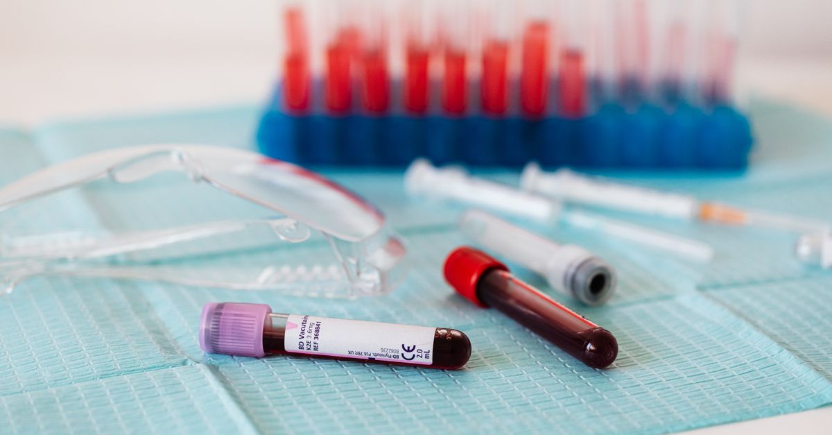
What Does CRP Actually Measure?
C-reactive protein (CRP) is a pentameric protein synthesized by the liver in response to inflammatory cytokines — primarily interleukin-6 (IL-6) and tumor necrosis factor-alpha (TNF-α). Named for its ability to bind the C-polysaccharide of Streptococcus pneumoniae, CRP was originally valued as an acute-phase reactant: a blunt instrument for detecting infection or tissue damage.
But that's only half the story.
In functional and preventive medicine, high-sensitivity CRP (hs-CRP) has emerged as one of the most clinically significant markers of low-grade, chronic inflammation — the kind that doesn't announce itself with a fever but quietly drives cardiovascular disease, insulin resistance, autoimmune conditions, and accelerated aging. Understanding what CRP actually measures — and what it doesn't — is the starting point for interpreting your labs with any real precision.
How CRP Is Produced
When immune cells detect cellular damage, infection, or metabolic stress, they release IL-6 into circulation. IL-6 travels to the liver, where hepatocytes rapidly ramp up CRP synthesis. CRP then circulates and binds to damaged or dying cells, apoptotic nuclei, and certain bacterial membranes, activating the complement cascade and facilitating phagocytosis. This is CRP doing its intended job: marking threats for immune clearance.
The problem is that the same pathway fires — more slowly and at lower amplitude — in response to visceral adiposity, oxidized LDL cholesterol, periodontal disease, sleep apnea, dysbiosis, and psychological stress. These stimuli produce a persistent, low-grade IL-6 signal that keeps CRP chronically elevated in the 1–10 mg/L range. That's the zone conventional labs often label 'normal' but functional clinicians treat as a zone of active risk.
CRP vs. hs-CRP: Why the Test You Order Matters
Standard CRP assays are calibrated for acute illness — they detect values above roughly 3–5 mg/L. High-sensitivity CRP (hs-CRP) uses a more sensitive immunoassay capable of detecting values as low as 0.1 mg/L, making it the preferred test for cardiovascular and metabolic risk stratification.
| Test | Detection Range | Primary Use Case |
|---|---|---|
| Standard CRP | ~3–500+ mg/L | Acute infection, post-surgical monitoring |
| High-sensitivity CRP (hs-CRP) | 0.1–10 mg/L | Cardiovascular risk, chronic inflammation |
For a standard wellness blood panel, always request hs-CRP specifically. If your results only show 'CRP: <5 mg/L,' you may have missed a meaningful signal at 2.8 mg/L.
Reference Range vs. Optimal Range
This is where interpretation diverges sharply between conventional and functional approaches.
| Category | hs-CRP (mg/L) | Interpretation |
|---|---|---|
| Optimal | < 0.5 | Low inflammatory burden |
| Low risk | 0.5 – 1.0 | Generally acceptable |
| Moderate risk | 1.0 – 3.0 | Elevated cardiovascular risk; investigate root cause |
| High risk | > 3.0 | Significant inflammation; warrants clinical evaluation |
| Acute illness | > 10 | Rule out acute infection or autoimmune flare before interpreting |
The landmark JUPITER trial (Ridker et al., NEJM 2008; PMID: 18997196) enrolled 17,802 healthy individuals with LDL below 130 mg/dL but hs-CRP ≥ 2.0 mg/L. Those treated with rosuvastatin showed a 44% reduction in major cardiovascular events — confirming that elevated hs-CRP predicts hard outcomes independently of LDL. This is precisely why hs-CRP above 1.0 mg/L warrants action even when your cholesterol panel looks clean.
---
What Does Fibrinogen Actually Measure — and How Does It Relate to CRP?
Fibrinogen is a soluble plasma glycoprotein synthesized by the liver that converts to fibrin during clot formation. Like CRP, fibrinogen is an acute-phase reactant — it rises in response to the same IL-6 signal that drives CRP elevation. This makes fibrinogen a useful companion marker when interpreting an inflammatory picture.
Where CRP reflects the speed and intensity of the inflammatory response, fibrinogen reflects the body's coagulation readiness. Persistently elevated fibrinogen (above 350–400 mg/dL) signals both increased clotting risk and ongoing systemic inflammation. A 2012 meta-analysis of 154,000 participants from 31 prospective studies found that fibrinogen levels in the top third of the population were associated with approximately a two-fold increased risk of coronary heart disease compared to the bottom third, independent of conventional risk factors (Fibrinogen Studies Collaboration, JAMA 2012; PMID: 22571587).
Functional interpretation: When both hs-CRP and fibrinogen are elevated simultaneously, the likelihood of underlying systemic inflammation — as opposed to a transient trigger — increases substantially. This dual elevation is common in metabolic syndrome, non-alcoholic fatty liver disease, and active autoimmune conditions.
Optimal fibrinogen range: 200–350 mg/dL
If fibrinogen consistently exceeds 400 mg/dL alongside elevated hs-CRP, it's worth investigating root causes of chronic inflammation with your healthcare provider.
---
What Do Thyroid Antibodies Actually Measure — and Why Do They Affect CRP?
Thyroid peroxidase antibodies (TPO-Ab) and thyroglobulin antibodies (TG-Ab) are immunoglobulins produced when the immune system mistakenly targets thyroid tissue. These antibodies are the diagnostic hallmark of Hashimoto's thyroiditis (autoimmune hypothyroidism) and Graves' disease.
So why are they relevant in a CRP discussion?
Because autoimmune thyroid disease is an inflammatory condition. Active antibody-mediated thyroid destruction triggers local and systemic cytokine release — including IL-6, which drives CRP upward. Studies have found that TPO-Ab-positive individuals with Hashimoto's display significantly higher hs-CRP levels than antibody-negative controls, even when TSH and free thyroid hormones remain within reference range (Baser et al., Thyroid 2015; PMID: 25646730).
This is a critical clinical nuance: a patient with subclinical Hashimoto's — normal TSH, elevated TPO-Ab, and hs-CRP of 2.4 mg/L — is displaying a coherent inflammatory picture. Treating CRP in isolation without identifying the antibody-mediated driver will produce limited results.
Optimal thyroid antibody ranges:
- TPO-Ab: < 9 IU/mL (many labs reference < 35; functional threshold is tighter)
- TG-Ab: < 1–4 IU/mL
If you've been told your thyroid antibodies are 'borderline,' request the actual numerical value. An antibody level of 32 IU/mL vs. 280 IU/mL tells very different stories about immune burden — and both may appear below the conventional cutoff at some labs.
---
What Does Free T4 Actually Measure — and Its Relationship to Inflammatory Markers?
Free T4 (thyroxine) is the unbound, biologically available form of the primary thyroid hormone produced by the thyroid gland. Unlike total T4, which includes protein-bound hormone, free T4 reflects what's actually accessible to cells. It is the dominant precursor that peripheral tissues convert into the metabolically active free T3.
From an inflammation standpoint, free T4 connects to CRP through two mechanisms:
- Hypothyroidism and inflammation: Low free T4 — whether from Hashimoto's, iodine insufficiency, or pituitary dysfunction — is associated with elevated inflammatory markers. Thyroid hormone helps regulate NF-κB, a master transcription factor controlling pro-inflammatory cytokine production. When thyroid output falls, NF-κB activity can rise, contributing to elevated hs-CRP (Kvetny et al., Thyroid 2004; PMID: 15186602).
- Thyroid hormone and CRP in cardiovascular risk: Subclinical hypothyroidism (normal TSH but low-normal free T4) has been independently associated with increased hs-CRP and cardiovascular risk in several prospective studies.
Functional free T4 optimal range: 1.1–1.4 ng/dL (most lab reference ranges span 0.8–1.8 ng/dL — a much wider window that obscures borderline insufficiency).
Always interpret free T4 alongside TSH and free T3. A free T4 sitting at 0.9 ng/dL is technically 'in range' but may reflect inadequate thyroid reserve in a symptomatic patient.
---
What Does Free T3 Actually Measure — and Why It's the CRP-Adjacent Marker Most Labs Skip
Free T3 (triiodothyronine) is the most biologically potent thyroid hormone. While the thyroid gland secretes some T3 directly, approximately 80% of circulating free T3 is converted peripherally from free T4 — primarily in the liver, kidneys, and gut. This conversion step is sensitive to inflammation, nutrient deficiencies (selenium, zinc, iron), and elevated cortisol.
The CRP–T3 connection is particularly important: chronic elevation of hs-CRP signals an inflammatory state that actively impairs T4-to-T3 conversion, effectively leaving cells thyroid-depleted even when free T4 looks adequate. This is the mechanism behind the well-documented phenomenon of 'low T3 syndrome' (also called 'euthyroid sick syndrome' or 'non-thyroidal illness syndrome') observed in chronically ill patients.
For a deeper look at how thyroid hormone conversion interacts with metabolic health, see our guide on thyroid optimization and lab testing.
Functional free T3 optimal range: 3.2–4.4 pg/mL (conventional labs often reference 2.3–4.2 pg/mL).
A patient with free T3 at 2.6 pg/mL and hs-CRP at 2.1 mg/L is displaying a coherent inflammatory suppression of thyroid conversion — not merely two unrelated findings.
---
What Causes CRP to Elevate Silently?
Beyond thyroid autoimmunity and conversion impairment, common drivers of subclinical hs-CRP elevation include:
- Visceral adiposity: Adipose tissue, especially visceral fat, is metabolically active and secretes IL-6 directly
- Dysbiosis and intestinal permeability: Lipopolysaccharide (LPS) translocation from gram-negative gut bacteria activates toll-like receptor 4 (TLR4), triggering hepatic CRP synthesis
- Oxidized LDL and metabolic syndrome: Small dense LDL particles are more inflammatory than large buoyant particles
- Sleep deprivation: Even short-term sleep restriction raises hs-CRP; a trial found that sleeping ≤6 hours per night was associated with significantly higher hs-CRP than sleeping 7–8 hours (Meier-Ewert et al., Journal of the American College of Cardiology 2004; PMID: 14975483)
- Periodontal disease: Oral bacteria trigger systemic IL-6 responses
- Psychological stress and HPA dysregulation: Elevated cortisol initially suppresses CRP, but chronic stress dysregulates this feedback, contributing to chronic low-grade elevation
---
What This Means for Your Formula
At Ones, every formula is built from an individual's actual data — blood work, wearable metrics, and health history — not a demographic category. When hs-CRP appears elevated alongside other relevant markers, the AI health practitioner identifies which upstream drivers are most likely active and selects ingredients accordingly.
For inflammatory profiles, several Ones ingredients are particularly relevant:
Omega-3 (EPA/DHA): EPA and DHA are the most rigorously studied natural modulators of hs-CRP. A meta-analysis of 68 randomized controlled trials found omega-3 supplementation significantly reduced CRP, IL-6, and TNF-α across diverse populations (Calder et al., British Journal of Nutrition 2010). Ones includes omega-3 at clinically meaningful EPA/DHA doses calibrated to your inflammatory burden.
Vitamin D3 + K2 (MK-7): Vitamin D deficiency is consistently associated with elevated hs-CRP. Vitamin D receptors are present on immune cells, and D3 supplementation has been shown to modulate NF-κB signaling and reduce inflammatory cytokine output. Ones pairs D3 with K2 (MK-7) to support vascular health alongside inflammation management.
Ones Liver Support Blend: Given that CRP is synthesized in the liver and that hepatic inflammation is a common upstream driver of elevated hs-CRP, the proprietary Liver Support blend targets hepatic cellular health as part of addressing systemic inflammatory load.
For autoimmune thyroid-driven inflammation patterns — where elevated TPO-Ab and low free T3 accompany elevated hs-CRP — the Thyroid Support blend and targeted selenium (selenomethionine) may be incorporated, given selenium's documented role in reducing TPO antibody titers (Gärtner et al., Journal of Clinical Endocrinology & Metabolism 2002; PMID: 11932302).
Formulas are delivered in either a 6- or 9-capsule daily plan, with the AI selecting the appropriate plan based on the breadth and complexity of your findings.
---
Key Takeaways
- CRP is an upstream, integrative marker — it doesn't pinpoint a single cause but signals that inflammatory cytokines are active. The driver could be metabolic, autoimmune, infectious, or behavioral.
- Optimal hs-CRP is below 1.0 mg/L, not the conventional 'below 3.0 mg/L' cutoff. Values between 1.0–3.0 mg/L represent a zone of meaningful cardiovascular and metabolic risk.
- Fibrinogen is a complementary acute-phase marker. Dual elevation of fibrinogen (>400 mg/dL) and hs-CRP strengthens the case for systemic, chronic inflammation.
- Thyroid antibodies, free T4, and free T3 connect directly to CRP biology. Autoimmune thyroid disease elevates CRP; elevated CRP impairs T4-to-T3 conversion — creating a bidirectional feedback loop.
- The hs-CRP test must be specifically ordered. Standard CRP misses the clinically significant 1–5 mg/L range where most functional intervention is warranted.
- Interpreting CRP in isolation is a mistake. Run it alongside the full thyroid panel (TSH, free T4, free T3, TPO-Ab, TG-Ab), fibrinogen, metabolic markers, and a lipid panel for a complete picture of your inflammatory biology.