Women's Health
Should Women Get Bone Density Tested?
One in two women over 50 will break a bone due to osteoporosis — yet most have no idea their bone density is declining until a fracture happens. Bone loss begins as early as your mid-30s, accelerates sharply around menopause, and is largely preventable when caught early. Here's what every woman should know about testing.
Should Women Get Bone Density Tested?
Yes — most women should get a baseline bone density scan by age 65 at the latest, and earlier if risk factors are present. Bone loss is clinically silent for decades, which means a fracture is often the first symptom. The exception is younger women with no risk factors, strong dietary calcium intake, and regular weight-bearing exercise, who can reasonably wait until perimenopause to begin discussions with their physician.
---
What Is a Bone Density Test and How Does It Work?
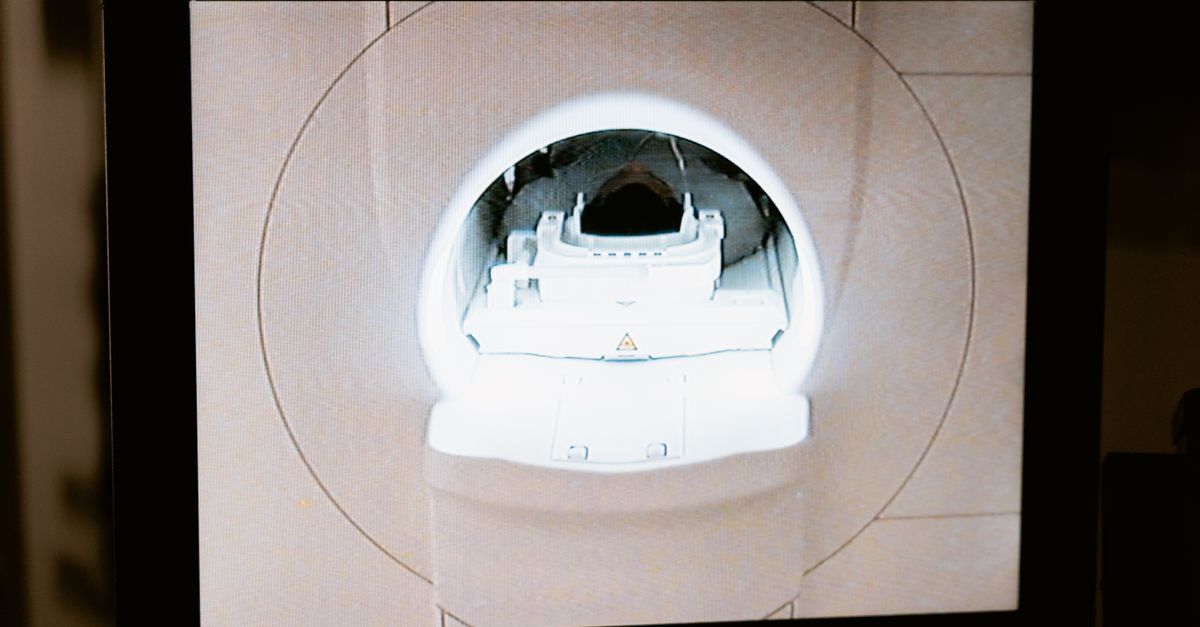
The standard tool is dual-energy X-ray absorptiometry, commonly called a DEXA scan. It measures bone mineral density (BMD) at the hip and lumbar spine, then expresses the result as a T-score — the number of standard deviations above or below the average BMD of a healthy young adult.
| T-Score Range | Classification |
|---|---|
| −1.0 and above | Normal |
| −1.0 to −2.5 | Osteopenia (low bone mass) |
| −2.5 and below | Osteoporosis |
The U.S. Preventive Services Task Force (USPSTF) recommends routine screening for all women aged 65 and older, and for younger postmenopausal women whose 10-year fracture risk equals or exceeds that of a 65-year-old Caucasian woman — typically calculated using the FRAX algorithm (USPSTF, 2018; doi.org/10.1001/jama.2018.7498).
FRAX incorporates age, sex, body weight, prior fracture history, smoking status, glucocorticoid use, rheumatoid arthritis diagnosis, and parental hip fracture history into a probabilistic risk estimate. A FRAX score of 20% or higher for major osteoporotic fracture, or 3% or higher for hip fracture specifically, is a common threshold for initiating pharmacological treatment.
Why Bone Loss Often Goes Undetected in Women
Estrogen is the primary regulator of bone remodeling in women. It slows osteoclast activity — the cells that break bone down — while supporting osteoblasts that build new bone. During the perimenopausal transition, estradiol levels fall rapidly, and bone turnover accelerates sharply. Women can lose 1–3% of their bone mass per year in the first five years after menopause, and up to 20% of total bone mass in the decade following their final period (Eastell et al., Nature Reviews Endocrinology 2016; PMID: 26857146).
Because this loss is painless and invisible, many women only discover it after a "fragility fracture" — a break from minimal trauma such as a fall from standing height or even a cough that cracks a rib. Hip fractures carry a one-year mortality rate approaching 20–25% in older women, making prevention a genuine survival issue, not merely a quality-of-life concern.
Understanding how estradiol levels relate to bone health is part of why comprehensive hormone testing matters alongside a DEXA scan. Similarly, tracking FSH levels as they rise in perimenopause can help women and their clinicians anticipate when bone protection should become a priority.
When Should Women Get a Bone Density Test?
The short answer depends on your risk profile:
- Age 65+: Routine screening regardless of symptoms or risk factors (USPSTF recommendation).
- Postmenopausal under 65: Test if FRAX score is elevated, or if you have a history of low-trauma fracture, prolonged steroid use, low body weight (BMI < 20), eating disorder history, or malabsorption conditions such as celiac disease.
- Premenopausal women: Testing is generally not recommended unless there are specific risk factors — hyperthyroidism, hyperparathyroidism, long-term use of medications that deplete bone (aromatase inhibitors, GnRH agonists, anticonvulsants), or a history of amenorrhea.
- Perimenopause (typically 45–55): A baseline scan is increasingly recommended by endocrinology societies because it establishes a reference point before the steepest period of loss begins.
Repeat testing intervals depend on baseline BMD: women with normal density and low risk may not need rescreening for 10–15 years, while those with osteopenia typically rescreen every 1–2 years.
Key Risk Factors That Move Your Testing Timeline Earlier
Several modifiable and non-modifiable factors accelerate bone loss:
- Low calcium and vitamin D intake — dietary calcium below 1,000 mg/day and serum 25(OH)D below 30 ng/mL impair bone mineralization
- Inadequate protein intake — collagen synthesis depends on dietary protein; low intake is independently associated with lower BMD (Sahni et al., Journal of Bone and Mineral Research 2010; PMID: 20200952)
- Sedentary lifestyle — weight-bearing and resistance exercise are the primary mechanical stimuli for bone remodeling
- Smoking and heavy alcohol use — both directly impair osteoblast function
- Low body weight — adipose tissue contributes to estrogen production postmenopausally; very lean women lose this secondary source
- Elevated cortisol — chronic psychological stress and elevated endogenous cortisol suppress bone formation; tracking cortisol is especially relevant for women under chronic stress
- Low iron and hemoglobin — iron deficiency and anemia have been associated with impaired bone metabolism; women at risk of low hemoglobin may face compounding risks
Nutrients That Directly Support Bone Mineral Density
Pharmacological treatment (bisphosphonates, denosumab, raloxifene) is reserved for women who already have osteoporosis or who meet high fracture-risk thresholds. For the majority of women — especially those in the osteopenia range or seeking prevention — nutrition and supplementation form the foundation of bone protection.
Vitamin D3 + K2 (MK-7)
Vitamin D3 enhances intestinal calcium absorption, but without adequate vitamin K2 (specifically the MK-7 isoform), calcium can be deposited in arterial walls rather than bone. A 2013 randomized controlled trial in postmenopausal women found that 180 mcg/day of MK-7 significantly increased bone strength indices and slowed the rate of bone loss compared to placebo over three years (Knapen et al., Osteoporosis International 2013; PMID: 23525894). The combination of D3 and MK-7 is now considered the evidence-based standard for bone-supportive supplementation.
Magnesium
Roughly 60% of the body's magnesium is stored in bone, where it influences hydroxyapatite crystal formation and parathyroid hormone activity. Magnesium deficiency has been associated with reduced osteocalcin levels and impaired vitamin D metabolism. Magnesium glycinate is a highly bioavailable form that avoids the laxative effect common with magnesium oxide, making it practical for daily supplementation.
Calcium
Adequate dietary calcium (1,000–1,200 mg/day depending on age) remains the bedrock of bone nutrition, though supplemental calcium beyond dietary needs should be approached carefully — large supplemental doses without K2 have raised cardiovascular concerns in some observational studies. Food-first calcium from dairy, leafy greens, and fortified foods is the preferred strategy.
How Ones Addresses This
Ones is a personalized supplement platform that uses blood work, wearable data, and health history to build a custom daily capsule formula. For women focused on bone health, the AI practitioner evaluates vitamin D status directly from lab results rather than applying a blanket dose — because the right amount of D3 to move serum 25(OH)D into the optimal 40–60 ng/mL range varies meaningfully from person to person.
When bone support is a priority, Ones formulas can include Vitamin D3 + K2 (MK-7) dosed to match the clinical evidence — the K2 component is specifically the MK-7 form at 180 mcg, mirroring the Knapen 2013 RCT protocol. Ones also includes Magnesium Glycinate as part of its Magnesium Complex blend, calibrated to avoid the tolerance issues of oxide forms. Where stress and cortisol dysregulation are flagged in a woman's data — an important secondary driver of bone loss — Ones may incorporate Ashwagandha (KSM-66, 600 mg), which has demonstrated significant reductions in serum cortisol in a 60-day double-blind RCT (Chandrasekhar et al., Indian Journal of Psychological Medicine 2012; PMID: 23439798).
Formulas are built in 6 or 9-capsule daily plans selected by the AI based on the totality of a user's findings — not by user preference — which means bone support ingredients are weighed against everything else flagged in the data.
A Note on Secondary Keywords in This Context
Some amino acids are occasionally discussed in relation to bone and musculoskeletal health, though the evidence is less direct than for calcium, vitamin D, and K2. L-glutamine plays a role in intestinal integrity — compromised gut lining impairs nutrient absorption including calcium and magnesium. L-carnitine has been studied in a limited way for bone density in postmenopausal women, with one trial suggesting it may partially counteract bone resorption (Maggio et al., Calcified Tissue International 2013; PMID: 23397298). Taurine contributes to antioxidant defense and may play a minor supporting role in bone matrix quality, though clinical evidence in humans remains preliminary. L-tyrosine is a thyroid hormone precursor, and given that thyroid abnormalities can accelerate bone loss, maintaining optimal thyroid function is indirectly relevant — but L-tyrosine supplementation is not a bone density intervention by itself. These amino acids are worth understanding in context, but they do not replace the foundational bone-support triad of calcium, vitamin D3, and vitamin K2.
Interpreting Your DEXA Results: What Happens Next
Receiving a DEXA result is not the end of the conversation — it's the beginning. Here is a practical framework for acting on results:
- Normal T-score (above −1.0): Reinforce calcium and vitamin D intake, maintain weight-bearing exercise, schedule next scan per your physician's guidance (typically 10–15 years if risk is low).
- Osteopenia (−1.0 to −2.5): Optimize nutritional status, add K2 and magnesium if not already supplementing, consider FRAX calculation to determine whether pharmacological treatment is warranted, and rescreen in 1–3 years.
- Osteoporosis (below −2.5) or prior fragility fracture: Work with a physician or endocrinologist on pharmacological options (bisphosphonates are first-line), continue nutritional optimization, and address fall-prevention strategies.
- High FRAX score despite normal BMD: BMD is only one input. A high fracture-risk score may still warrant treatment because cortical microarchitecture and trabecular quality are not fully captured by DEXA.
Key Takeaways
- Women should begin discussing bone density testing with their physician by perimenopause and get a formal DEXA scan by age 65 at the latest — earlier if any significant risk factors are present.
- Bone loss is clinically silent; a fragility fracture is often the first sign, making proactive testing genuinely life-altering.
- Estrogen decline at menopause drives 1–3% annual bone loss; monitoring estradiol and FSH levels provides early warning.
- The evidence-based nutritional foundation for bone protection is calcium (food-first), Vitamin D3 calibrated to serum levels, and Vitamin K2 as MK-7 (180 mcg/day has the strongest RCT support).
- Chronic cortisol elevation is an underappreciated driver of bone loss; managing stress — and testing cortisol — belongs in a comprehensive bone health plan.
- Personalized supplement formulas built from your actual lab data, like those from Ones, can target the specific nutrient gaps most relevant to your bone density risk rather than applying generic doses.