Supplements
What Happens to AMH Levels During Pregnancy?
AMH levels behave in a surprising and counterintuitive way during pregnancy — they fall significantly in the first trimester, even in women with robust ovarian reserves before conception. Understanding this pattern is critical if you've had AMH tested while pregnant or shortly after delivery, because a single low reading can be deeply misleading without context.
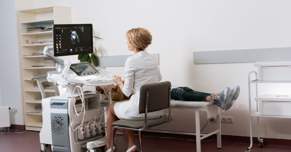
What Happens to AMH Levels During Pregnancy?
AMH drops sharply — often by 50–70% — during the first trimester of pregnancy, then continues to decline through the second trimester before partially recovering postpartum. This is a normal physiological pattern, not a sign that your ovarian reserve is being depleted by pregnancy. The important exception: women who entered pregnancy with genuinely low AMH may see levels fall into an undetectable range, which is worth discussing with a reproductive endocrinologist after delivery.
---
Why AMH Falls So Dramatically in Early Pregnancy
Anti-Müllerian hormone is produced almost exclusively by granulosa cells in small antral and preantral follicles in the ovaries. Its serum concentration is widely used as a proxy for ovarian reserve — the quantity of egg-containing follicles remaining. Outside of pregnancy, AMH is remarkably stable across the menstrual cycle, which is one reason it became the preferred ovarian reserve marker over FSH or antral follicle count.
During pregnancy, however, the hormonal environment shifts dramatically. Rising human chorionic gonadotropin (hCG) and progesterone suppress follicle recruitment. When fewer small follicles are actively developing, the granulosa cells producing AMH become less active, and circulating AMH falls as a result (La Marca et al., Journal of Clinical Endocrinology & Metabolism 2005; PMID: 15687341).
A prospective cohort study by Tremellen and Savulescu (2014; PMID: 24768706) tracked AMH in women from before conception through delivery and found that median AMH fell by approximately 60% by the end of the first trimester. This reduction was consistent regardless of whether baseline AMH was high, normal, or borderline low. The same study documented that AMH began recovering within six weeks postpartum in most participants, supporting the interpretation that this is a reversible suppression rather than true follicle loss.
For a deeper look at how AMH fits into the broader hormonal picture, see what Anti-Müllerian Hormone tells you about your hormones — it covers the clinical reference ranges and what patterns actually predict fertility outcomes.
---
How Low Can AMH Go During Pregnancy, and Is This Harmful?
In practical terms, AMH can fall to levels that, outside of pregnancy, would indicate severely diminished ovarian reserve. Values below 0.5 ng/mL — sometimes below the limit of detection of standard assays — have been documented in otherwise healthy pregnant women who had normal preconception AMH (Köninger et al., Reproductive Biology and Endocrinology 2019; PMID: 31856836).
This is not harmful to the pregnancy or to future fertility. The follicles themselves are not being destroyed. The granulosa cells are simply in a quiescent state because the pregnancy hormones have suppressed the normal recruitment signaling. Think of it like a factory that has temporarily reduced production — the machinery is intact.
The clinical danger is misinterpretation. A woman who has AMH tested during an early pregnancy (particularly one that ends in miscarriage and is later identified) or shortly after delivery — before levels have recovered — may receive a reading that appears to indicate dramatically reduced ovarian reserve. Clinicians who are unaware of the pregnancy status at the time of the draw may counsel incorrectly about future fertility prospects.
If you've received a surprising AMH result, it's worth understanding the full panel of hormones in context. The article on what cholesterol tells you about your hormones illustrates how single-marker readings are routinely misread without a systems-level view — the same principle applies here.
---
The Postpartum Recovery: How Long Does It Take for AMH to Rebound?
The timeline for AMH recovery after delivery is not fully standardized in the literature, but the available evidence is reasonably consistent. Most studies find that AMH returns to approximately pre-pregnancy levels within three to six months postpartum, though breastfeeding may modestly delay this recovery.
A study by Müller and colleagues (2020; doi: 10.1007/s10815-020-01717-0) followed 47 women from preconception through 12 months postpartum and found that:
- AMH at 12 weeks gestation was roughly 55% of baseline
- AMH at 20 weeks was the nadir, averaging 47% of baseline
- AMH at six weeks postpartum had recovered to approximately 80% of baseline
- AMH at six months postpartum was statistically indistinguishable from preconception values
Women who were exclusively breastfeeding showed slightly slower recovery, likely because lactation-associated prolactin elevations further suppress gonadotropin activity and follicle stimulation. This is an important consideration for women who plan to conceive again shortly after a first pregnancy — testing AMH before six months postpartum, especially while nursing, may still underestimate true reserve.
---
Does Pregnancy Affect AMH Differently Based on Age or Baseline Reserve?
This is one of the more clinically nuanced questions in reproductive endocrinology. The short answer is: yes, but the proportional decline appears similar regardless of baseline.
Women with high preconception AMH (for example, those with polycystic ovary syndrome who often have AMH values three to five times the age-adjusted average) show the largest absolute decline in numbers, but because they started higher, they remain well above detection limits throughout pregnancy. Their postpartum return is rapid and complete.
Women with low preconception AMH — say, under 1.0 ng/mL — experience the same proportional suppression but may fall below the sensitivity threshold of standard assays during pregnancy. This creates what looks like an undetectable AMH on paper, which can be alarming if the context of pregnancy is not taken into account.
Age compounds this. An older woman with borderline reserve who becomes pregnant may have AMH levels that appear consistent with premature ovarian insufficiency if tested during the second trimester. The Tremellen cohort (PMID: 24768706) found that women over 38 with AMH under 1.5 ng/mL preconception were disproportionately likely to show undetectable AMH by mid-pregnancy.
For related context on how hormonal markers shift across the reproductive lifespan, the piece on what SHBG tells you about your hormones is worth reading alongside this one — SHBG, like AMH, is heavily influenced by pregnancy and is similarly prone to misinterpretation when not contextualized.
---
AMH and Pregnancy Loss: What the Research Actually Shows
One of the more emotionally charged questions surrounding AMH in pregnancy is whether low levels — whether pre-existing or discovered during workup after a loss — predict miscarriage risk. The evidence here deserves a careful reading.
AMH does not appear to be a direct predictor of miscarriage risk in natural conceptions. A large meta-analysis by Depmann and colleagues (Human Reproduction Update 2016; PMID: 26758258) examined data from over 30 studies and found no statistically significant association between preconception AMH and miscarriage rate in women who conceived naturally. Low AMH predicts difficulty conceiving — specifically, fewer available eggs — but not a higher rate of chromosomal or developmental problems in the eggs that do ovulate.
In assisted reproductive technology (ART) cycles, the picture is more complex. Low AMH is associated with poor ovarian response to stimulation, which reduces the number of embryos available for transfer. Fewer embryos mean fewer chances, but the quality of embryos retrieved from low-AMH women is not necessarily inferior to those from high-AMH women of the same age — age itself is the dominant determinant of embryo quality.
If you're navigating a recurrent pregnancy loss workup, a comprehensive hormonal panel including thyroid function, cortisol, and iron status is often more diagnostically relevant than AMH alone. The article on what iron tells you about your hormones covers how iron deficiency — which is extremely common in the periconception period — interacts with hormone signaling in ways that are frequently overlooked.
---
When Should You Actually Test AMH During or After Pregnancy?
The clinical consensus is straightforward: do not use AMH tested during pregnancy to draw conclusions about ovarian reserve. The value is suppressed physiologically and not interpretable in the same framework used for non-pregnant women.
The ideal timing for AMH testing to assess ovarian reserve is:
- Before conception, as part of a preconception panel
- At least six months postpartum (and ideally after weaning, if breastfeeding)
- After any evaluation for secondary infertility, with the date of last delivery and current breastfeeding status noted on the lab requisition
If you do receive an AMH result that was drawn during pregnancy — perhaps as part of a workup after a loss — treat it as uninterpretable for reserve purposes. Request a repeat draw at the appropriate postpartum interval before making any clinical decisions based on that number.
This timing awareness matters because decisions about fertility preservation (egg freezing), donor egg consideration, or aggressive IVF protocols can all be influenced by AMH — and getting them wrong in either direction has real consequences.
---
What This Means for Your Formula
AMH itself is not a target of supplementation — no ingredient raises AMH directly, and any product claiming otherwise is not supported by clinical evidence. However, the underlying biology that AMH reflects — follicle health, mitochondrial function in granulosa cells, and the hormonal environment supporting ovarian function — is influenced by several well-studied nutrients.
For women who are concerned about ovarian reserve and overall reproductive hormonal health, two ingredients in the Ones catalog are directly relevant:
- CoQ10/Ubiquinol (200mg): Mitochondrial function in oocytes and granulosa cells is energy-intensive and declines with age. CoQ10 has been studied specifically in the context of ovarian response, with a randomized trial by Xu et al. (Journal of Ovarian Research 2018; PMID: 29558944) showing that 600mg/day CoQ10 for 60 days before IVF improved ovarian response parameters in poor responders. Ones includes ubiquinol — the active, reduced form — at a clinically relevant dose calibrated to each user's profile.
- Zinc: Zinc is essential for follicle maturation, ovulation signaling, and early embryonic development. Deficiency is associated with impaired reproductive function. Ones includes zinc at doses informed by individual lab data and dietary intake — not a blanket RDA — which matters because zinc toxicity from oversupplementation is a real risk in women who are already eating zinc-rich diets. For more on the hormonal role of zinc, see what zinc tells you about your hormones.
Ones doesn't offer AMH-specific formulas because AMH is a biomarker, not a deficiency. What Ones does is analyze the full picture — including lab values, wearable data, and health history — to address the modifiable factors that support reproductive hormonal health, rather than chasing a single number.
---
Key Takeaways
- AMH falls by 50–70% during pregnancy — this is normal physiology, not follicle loss, and reverses postpartum.
- The nadir typically occurs in the second trimester; recovery to baseline usually takes three to six months after delivery, and may be slower while breastfeeding.
- Do not interpret an AMH result drawn during pregnancy as a measure of true ovarian reserve — it will almost always underestimate your actual reserve.
- Women with already-low preconception AMH may see levels drop to undetectable ranges during pregnancy; this is still not clinically meaningful in isolation.
- AMH does not predict miscarriage risk in natural conceptions — low AMH affects quantity of available eggs, not their quality.
- CoQ10 and zinc are the two best-studied nutrients in the context of granulosa cell and oocyte health; both require individualized dosing rather than blanket supplementation.
---
This article is for informational purposes only and does not constitute medical advice. Consult a reproductive endocrinologist or licensed healthcare provider before making decisions about fertility testing, treatment, or supplementation.